CASE 84
1. What are potential causes of this appearance? (Choose all that apply.)
B. Lymphoma
C. Angiosarcoma
D. Lung cancer
2. Which vessel is most severely narrowed?
A. Azygos vein
3. What is the most likely diagnosis?
B. Lymphoma
C. Angiosarcoma
D. Lung cancer
4. What is the most appropriate treatment in the acute setting?
A. Chemotherapy
C. Stent
D. Surgery
ANSWERS
References
Sheth S, Ebert MD, Fishman EK. Superior vena cava obstruction evaluation with MDCT. AJR Am J Roentgenol. 2010;194(4):W336–W346.
Wilson LD, Detterbeck FC, Yahalom J. Clinical practice Superior vena cava syndrome with malignant causes. N Engl J Med. 2007;356(18):1862–1869.
Comment
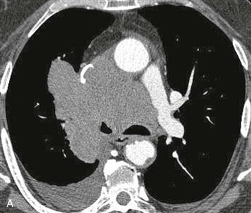
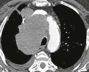
Clinical Presentation and Etiology
Signs and symptoms of superior vena cava syndrome are facial fullness and flushing, headache, upper extremity edema, and prominence of veins in the face and upper chest. The acute manifestation can be life-threatening. Most cases occur secondary to bronchogenic cancer. Other causes include fibrosing mediastinitis (most commonly secondary to histoplasmosis), lymphoma, other malignant tumors, and superior vena cava thrombosis.


