CASE 83
History: A patient presents with chest pain.
1. What should be included in the differential diagnosis? (Choose all that apply.)
A. Metastasis
B. Lipoma
C. Myxoma
D. Angiosarcoma
2. What is the most common mass in the heart?
A. Thrombus
B. Myxoma
C. Metastasis
D. Angiosarcoma
A. Left atrium
C. Right atrium
4. If this is a primary tumor, what is the most likely diagnosis?
A. Myxoma
B. Angiosarcoma
D. Rhabdomyoma
ANSWERS
Reference
Randhawa K, Ganeshan A, Hoey ET. Magnetic resonance imaging of cardiac tumors: part 2, malignant tumors and tumor-like conditions. Curr Probl Diagn Radiol. 2011;40(4):169–179.
Cross-Reference
Cardiac Imaging: The REQUISITES, ed 3, pp 281–282.
Comment
Pathology, Etiology, and Treatment
Approximately 98% of cardiac tumors are secondary tumors. The most common malignant primary cardiac tumor is angiosarcoma. Other primary malignant tumors are rare and include rhabdomyosarcoma, leiomyosarcoma, liposarcoma, and lymphoma. Definitive diagnosis is made with endomyocardial or open biopsy.
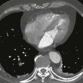
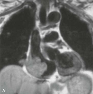
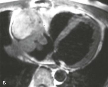
MRI
MRI can show tumor size and location and cardiac function. Contrast-enhanced sequences can delineate tumor margins and invasion into adjacent structures. The presence of enhancement does not signify malignancy; benign tumors enhance too. Features that suggest a primary malignant cardiac tumor include irregular or ill-defined margination, invasiveness, extension outside the heart (Figs. A and B), involvement of more than one chamber, central necrosis, large pericardial effusion, and lung nodules—which raise the possibility of metastases.