CASE 82
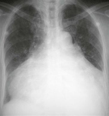
History: A patient presents with dyspnea.
1. What should be included in the differential diagnosis? (Choose all that apply.)
2. What is this appearance called?
3. What is the most likely diagnosis?
4. Which imaging modality best assesses the valve leaflets?
B. CT scan
C. MRI
D. Angiography
ANSWERS
Reference
Walker CM, Reddy GP, Steiner RM. Radiology of the heart. In: Rosendorff C, ed. Essential Cardiology. ed 3 New York: Springer; 2013.
Cross-Reference
Cardiac Imaging: The REQUISITES, ed 3, pp 205–206.
Comment
Pathology, Etiology, and Treatment
Tricuspid regurgitation can occur when there is an abnormality of one or more components of the valve apparatus: the anulus, leaflets, chordae, papillary muscles, and right ventricular wall. Tricuspid regurgitation can be acquired or congenital. Acquired causes include pulmonary hypertension secondary to mitral valve disease (most common cause), papillary muscle rupture, rheumatic heart disease, bacterial endocarditis, and carcinoid syndrome. The most common congenital cause is Ebstein anomaly. When tricuspid regurgitation is secondary to mitral valve disease, treatment of the mitral disease can relieve the tricuspid regurgitation. In more severe cases, tricuspid valve replacement or annuloplasty is performed.
Imaging
Chest radiographs demonstrate marked enlargement of the right atrium and ventricle (Figure). The heart can become massively enlarged, resulting in the so-called wall-to-wall heart. The differential diagnosis includes tricuspid regurgitation, dilated cardiomyopathy, and pericardial effusion. Echocardiography is performed to evaluate regurgitation further. Angiography and MRI can also be performed in selected cases. MRI is highly accurate for quantification of the severity of regurgitation.


