Procedure 80 Corrective Osteotomy of Malunited Distal Radius Fractures
Indications
 Deformity by itself may or may not be symptomatic. A decision for surgery should be based on functional impairment, pain, loss of motion, and limitation of grip strength.
 Alteration of radiocarpal alignment will affect normal carpal kinematics and changes in the vectors of the extensor and flexor tendons (Fig. 80-1).
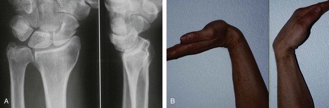
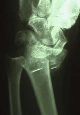
 Dorsally malunited fracture can result in a midcarpal instability (Figs. 80-2 and 80-3).


Examination/Imaging

Imaging
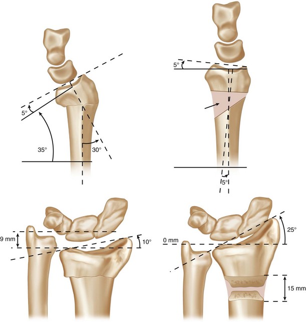
 Standard anteroposterior, lateral, and oblique radiographs of both wrists are essential for preoperative planning.
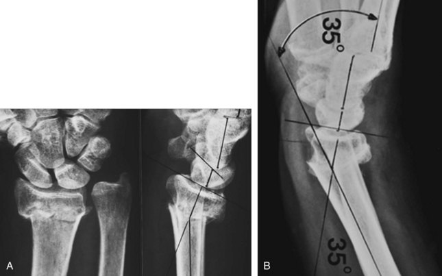
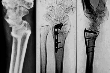
 For most deformities, tracings can be made from the anteroposterior and lateral radiographs and superimposed on each other. The angle and precise location of the deformity can be identified in the frontal and sagittal planes (Fig. 80-4).
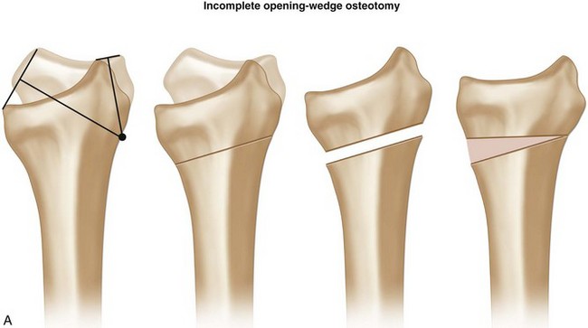
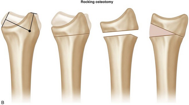
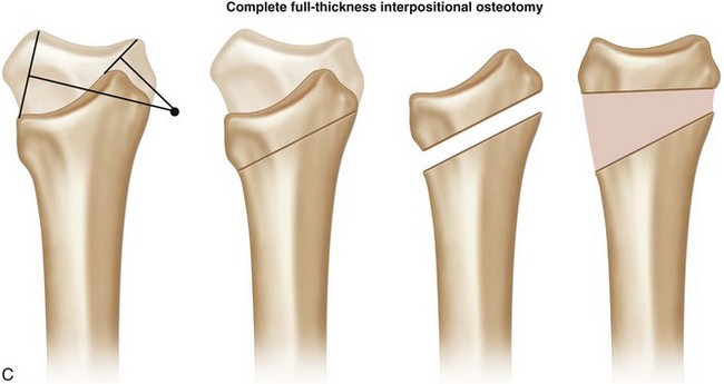
 By superimposing the tracings in the sagittal and frontal planes and drawing a line from the most volar and dorsal lips, a perpendicular line can be drawn midpoint on each line. Where these two intersect will define the type of osteotomy required and the need for lengthening of the distal fragment (Fig. 80-5).


Exposures
Volar
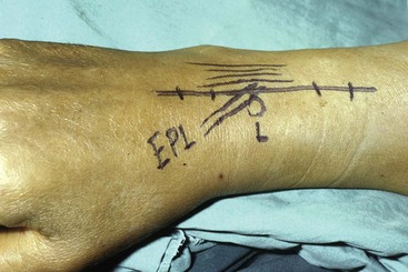
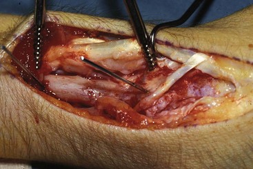
 The distal part of the anterior Henry approach involves exposure between the flexor carpi radialis and radial artery (Fig. 80-6).
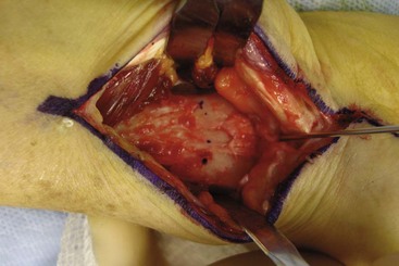
 The muscle belly of the flexor pollicis longus is retracted ulnarly to expose the pronator quadratus, which is elevated from its insertion on the radius (Fig. 80-7).

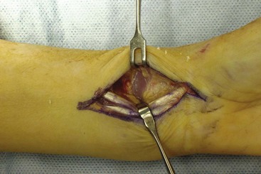
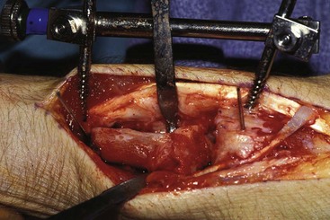
 Small Hohmann retractors are placed along the borders of the distal radius at the site of the deformity (Fig. 80-8).


Procedure
Volar Approach—Dorsal Malunion
Step 1
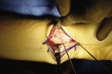
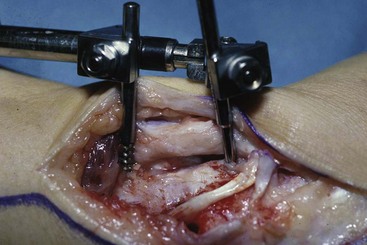
 The radiocarpal joint is located, and a smooth K-wire is placed parallel to the articular surface.
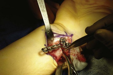
 Two additional K-wires are placed perpendicular to both the distal fragment and radial shaft (Fig. 80-11).
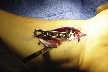
 The distal limb of the volar plate is temporarily screwed to the distal fragment with the proximal limb of the plate subtending the deformity angle in the sagittal and frontal planes (Fig. 80-12).
Step 2
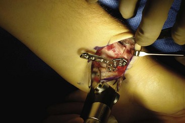
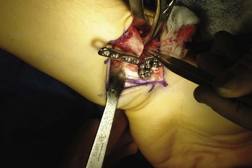
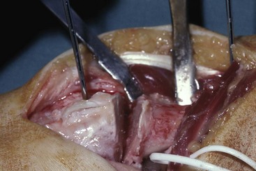
 The osteotomy cut is made parallel to the articular surface in the sagittal plane (Figs. 80-13 and 80-14).
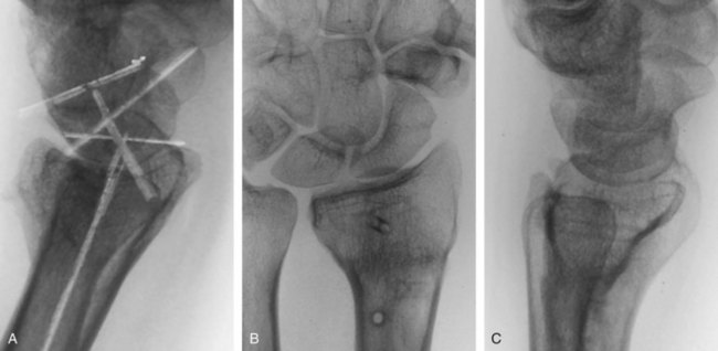
 The plate is reattached to the distal fragment (Fig. 80-15).
Step 3
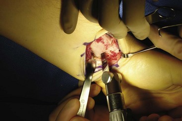
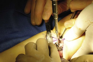
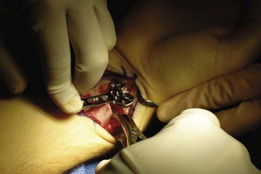
 By supinating the distal fragment while pronating the radial shaft, the dorsal callous and periosteum are released (Fig. 80-16).
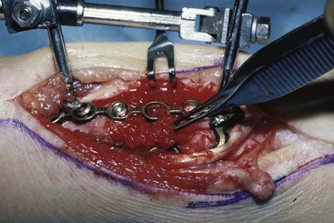
 A laminar spreader is placed in the osteotomy to help regain some length and help reduce the distal fragment (Fig. 80-17).


Postoperative Care and Expected Outcomes


Procedure


Procedure
Dorsal Approach
Step 1
 A smooth Kirschner wire is inserted through the dorsal wrist capsule parallel to the articular surface.
 Two threaded 2.5-mm Kirschner wires are inserted on both sides of the deformity to subtend the angle of correction (Fig. 80-22).
 A distractor can be attached to the pins to help gain length and maintain the corrected alignment (Fig. 80-23).
Step 1 Pearls
A small distractor is helpful to slowly gain length and realign the distal fragment (Fig. 80-23).
Placement of the K-wires follows the orientation determined by the preoperative plan.
If angular correction alone is required, the osteotomy is made parallel to the joint surface (Fig. 80-24).
If rotational correction is also required, the osteotomy should be made perpendicular to the distal fragment in both the frontal and sagittal planes.
Step 2
 Using a thin blade, the osteotomy is made parallel to the distal K-wires.
 The distractor can facilitate slow lengthening (Fig. 80-25).
Step 3
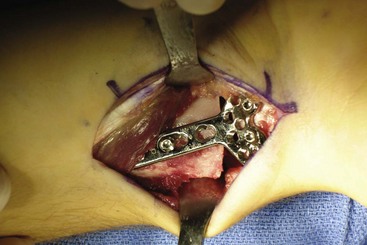
 Implant selection can be either two small 2.4-mm plates, one dorsally on the intermediate column and one between the first and second extensor compartments or, alternatively, an implant shaped to the dorsal anatomy of the distal radius.


Jupiter JB, Ring D. A comparison of early and late reconstruction of malunited fractures of the distal end of the radius. J Bone Joint Surg [Am]. 1996;78:739-748.
Prommersberger KJ, van Schoonhaven J, Lanz UB. Outcome after corrective osteotomy for malunited fractures of the distal end of the radius. J Hand Surg [Br]. 2002;27:55-60.
Ring D, Roberge C, Morgan T, et al. Osteotomy for malunited fractures of the distal radius: a comparison of structural and nonstructural autogenous bone grafts. J Hand Surg [Am]. 2002;27:216-222.


