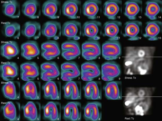
Case 8
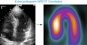
Echocardiography
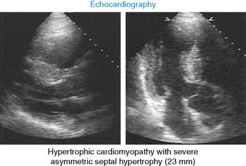
Hypertrophic cardiomyopathy (HCM) with severe asymmetric septal hypertrophy (23 mm).
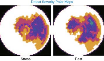
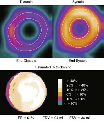
Teaching Points: MPI in Hypertrophic Cardiomyopathy
1. Quantitative perfusion polar maps compare regional tracer activity to a “normal” database, and patients with HCM will appear to have perfusion defects in myocardial regions with less hypertrophy (relative to regions with more severe hypertrophy).
2. Myocardial regions with severe hypertrophy will demonstrate an apparent reduction of regional systolic wall thickening due to the decreasing impact of the “partial volume effect” (relationship between wall thickness and apparent tracer activity).


