Procedure 74 Lunotriquetral Ligament Reconstruction Using a Slip of the Extensor Carpi Ulnaris Tendon
 See Video 55: Lunotriquetral Ligament Reconstruction Using the Extensor Carpi Ulnaris Tendon

Examination/Imaging
Clinical Examination
 Patients with LT instability present with ulnar-sided wrist pain. Other causes of ulnar-sided wrist pain include ulnar impaction syndrome, extensor carpi ulnaris (ECU) subluxation, distal radioulnar joint (DRUJ) instability, triangular fibrocartilage complex (TFCC) injuries, pisotriquetral arthritis, and hook of hamate fractures.

Imaging
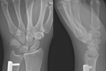
 A posteroanterior radiograph may show a step-off between the lunate and triquetrum and the disruption of the normal convex arc of the proximal carpal row (Gilula line). Unlike the scapholunate dissociation, a gap in the LT interval is rarely seen in patients with LT instability.
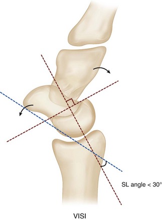
 Plain radiographs may show a volar intercalated segment instability (VISI) deformity, marked by volar tilting of the lunate (Fig. 74-3).
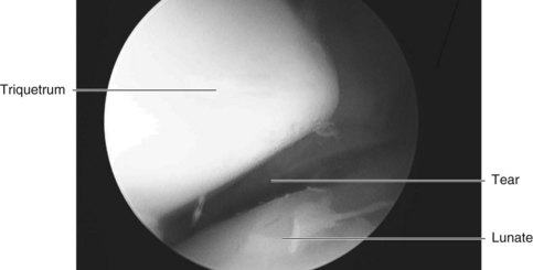
 Wrist arthroscopy is useful to diagnose an LT ligament tear when plain radiographs are normal (Fig. 74-4). The LT interosseous space is tightly closed in normal subjects, whereas in LT dissociations, the space can be opened with the probe, and a gap can be seen between the involved carpals from the midcarpal portal.
Surgical Anatomy
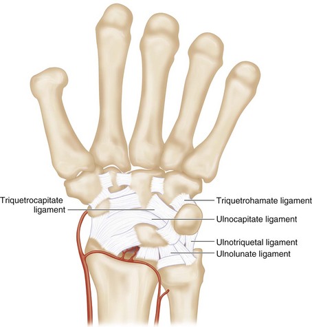
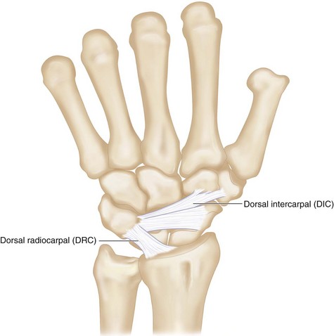
 The LT ligament is C shaped, spanning the (1) dorsal, (2) proximal, and (3) palmar edges of the joint surfaces. The palmar region is the thickest and strongest. It is the primary stabilizer of the LT joint. Additional supports of the LT joint include the ulnocarpal (ulnolunate, ulnocapitate, and ulnotriquetral ligaments), midcarpal (triquetrohamate and triquetrocapitate ligaments), and dorsal carpal (dorsal intercarpal and radiotriquetral) ligaments (Figs. 74-5 and 74-6).

Exposures
Dorsal
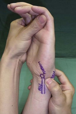
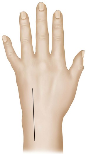
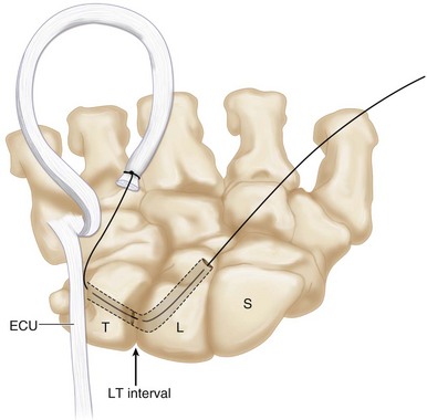
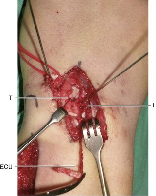
 A 6-cm longitudinal incision centered over the radiocarpal joint is made over the dorsal wrist between the fourth and fifth compartments (Fig. 74-7).

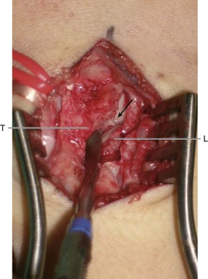
 Figure 74-8 shows a Freer elevator in the LT interval demonstrating a complete rupture of the LT ligament.
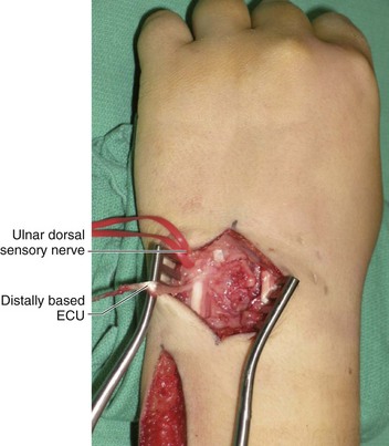
 The ECU tendon is identified in the proximal portion of this incision.
Pitfalls
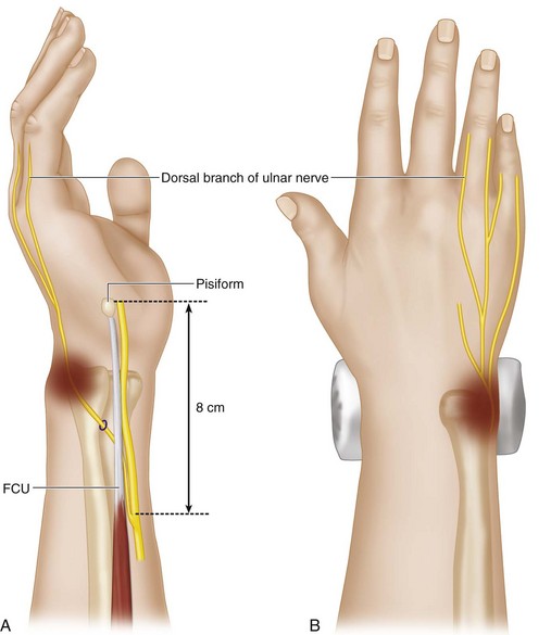
Branches of the ulnar dorsal sensory nerve should be protected. This nerve arises from the ulnar aspect of the ulnar nerve about 8 cm proximal to the pisiform. It then passes dorsal to the flexor carpi ulnaris (FCU) and pierces the deep fascia about 5 cm from the pisiform. It reaches the dorsum of the hand after coursing in close relation to the ulnar styloid process (Fig. 74-9).
Procedure

Step 2: Harvest a Strip of Distally Based ECU


Step 3: Reduction of Lunate and Triquetrum
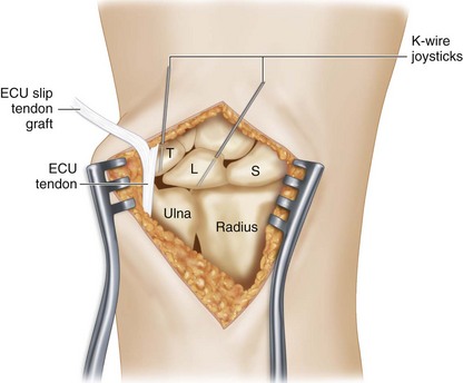
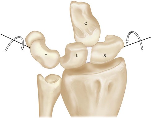
 Two 0.045-inch Kirschner wires (K-wires) are inserted into the lunate and the triquetrum to act as joysticks to reduce the carpal bones (Fig. 74-11).
Step 3 Pearls
The joystick K-wires are used to correct the VISI deformity. The lunate is flexed and the triquetrum extended (Fig. 74-12). The lunate K-wire should be inserted in an oblique distal to proximal direction, whereas the triquetral K-wire should be inserted in an oblique proximal to distal direction. The VISI deformity can be corrected by making both K-wires perpendicular to the axis of the forearm.
Step 4: Create a Bone Tunnel within Triquetrum and Lunate
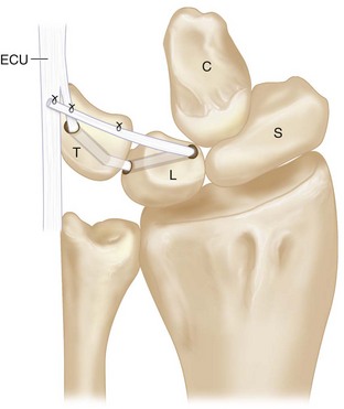
 A K-wire is passed from the distal, dorsal, and ulnar aspect of the triquetrum to exit at the proximal, palmar, and radial corner of triquetrum. Once acceptable placement of the K-wire is confirmed, a 2.5-mm cannulated drill is inserted over the K-wire to create a bone tunnel within the triquetrum.
 A similar bone tunnel is created from the distal, dorsal, and radial corner of the lunate to exit at the proximal, palmar, and ulnar aspect of the lunate (Fig. 74-13). A microcurette is used to remove the cartilage remnants at the exit sites of the drill holes.
Step 5: Pass the ECU through the Lunate and Triquetrum Tunnel

Step 6: Secure the ECU Graft to Itself and over the Remnant of the LT Ligament
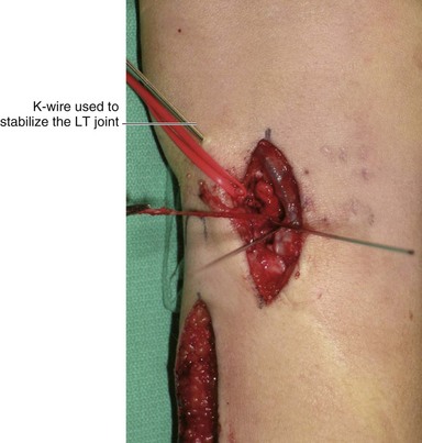
 The VISI deformity is corrected using the previously placed joystick wires, and the corrected position is maintained by a 0.045-inch K-wire passed between the lunate and triquetrum (Fig. 74-15).
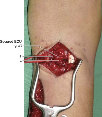
 The end of the tendon graft is sutured back to itself and to any remnants of the LT ligament over the triquetrum using 3-0 Ethibond sutures (Fig. 74-16). This will tighten the triquetrum against the lunate (Fig. 74-17).