CASE 74
1. What should be included in the differential diagnosis? (Choose all that apply.)
A. Transposition of great arteries
C. Pulmonary atresia with restrictive ventricular septal defect
2. What is the most likely diagnosis?
A. Hypoplastic left heart syndrome
B. Transposition of great arteries
3. What is the usual clinical presentation of this condition in neonates (<2 months old)?
A. Cyanosis
C. Heart failure with poor feeding and failure to thrive
4. Maternal ingestion of which drug during pregnancy is most commonly associated with Ebstein anomaly?
A. Atorvastatin
B. Lithium
C. Atenolol
D. Warfarin
ANSWERS
Reference
Ferguson EC, Krishnamurthy R, Oldham SA. Classic imaging signs of congenital cardiovascular abnormalities. Radiographics. 2007;27(5):1323–1334.
Cross-Reference
Cardiac Imaging: The REQUISITES, ed 3, pp 206–207.
Comment
Overview
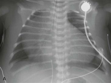
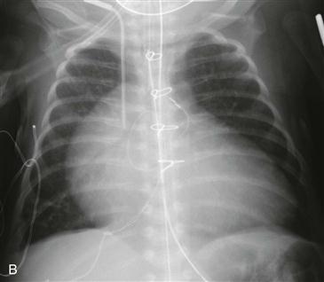
Ebstein anomaly is a rare congenital heart disease that usually results in cyanosis. This anomaly is characterized by ventricular displacement of the tricuspid valve leaflets resulting in a common ventriculoatrial chamber and often severe tricuspid regurgitation. Over time, there is progressive dilation of all right-sided chambers, which leads to increased tricuspid regurgitation and further chamber dilation. Most patients with Ebstein anomaly also have an atrial septal defect or patent foramen ovale. As right-sided pressures increase, a right-to-left shunt develops leading to cyanosis. This entity is characterized by normal to decreased pulmonary vasculature and massive right atrial enlargement, which may fill the entire right hemithorax. The aorta and pulmonary artery are typically small, leading to the characteristic box-shaped heart (Figs. A and B).
Associations
Ebstein anomaly is associated with an atrial septal defect or patent foramen ovale in approximately 90% of patients. Less common associations include supraventricular tachycardia (25% to 50%), pulmonary atresia or stenosis (25%), and Wolff-Parkinson-White syndrome (10%).
Imaging
A frontal radiograph shows massive cardiac enlargement (so-called wall-to-wall heart) and decreased pulmonary vasculature (Fig. A). Ebstein anomaly is the major consideration in a cyanotic patient. Much less common causes of this radiographic appearance include severe pulmonic stenosis with restrictive atrial septal defect, tricuspid atresia with a restrictive atrial septal defect, and pulmonary atresia with a restrictive ventricular septal defect.


