CASE 73
History: A 43-year-old woman presents with a rash.
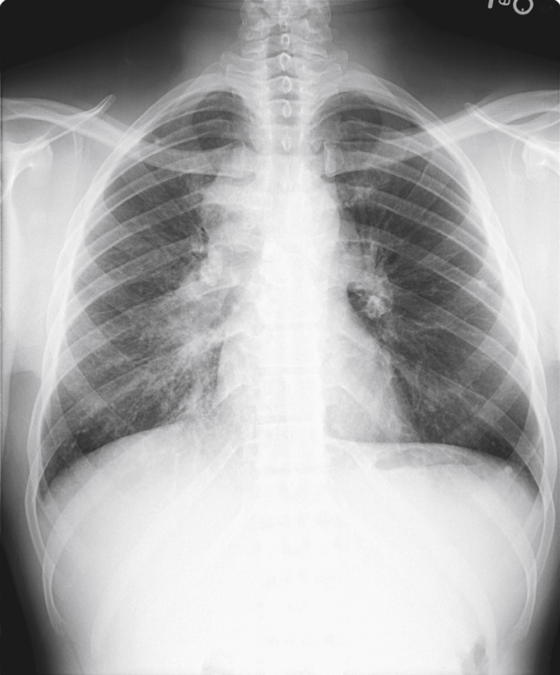
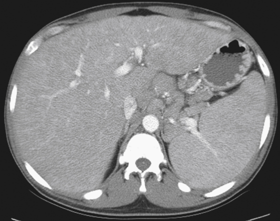
1. Which of the following should be included in the differential diagnosis of the imaging finding shown in the figures? (Choose all that apply.)
2. What is the most common intra-abdominal site of involvement by sarcoidosis?
3. Which of the following statements about abdominal sarcoid is true?
B. The most common imaging abnormality in abdominal sarcoidosis is extensive lymphadenopathy.
C. Hepatic involvement by sarcoid usually leads gradually but progressively to portal hypertension.
D. Biliary involvement by sarcoidosis results in strictures of the extrahepatic bile ducts.
4. Less than 10% of patients with sarcoidosis have gastrointestinal tract involvement. What places in the gut are most commonly involved?
ANSWERS
CASE 73
Sarcoidosis with Hepatosplenomegaly
1. A, B, C, and D
2. A
3. D
4. A
References
Farman J, Ramirez G, Rybak B, et al: Gastric sarcoidosis. Abdom Imaging. 1997;22:248–252.
Cross-Reference
Gastrointestinal Imaging: THE REQUISITES, 3rd ed, p 61.
Comment
Sarcoidosis is a disease generally seen involving the thorax, skin, or eyes, but it is also seen, on occasion, involving both solid and hollow viscera of the abdomen (see figures). The disease is of unknown etiology; at times correlation with some microorganism has been suggested but never proved. It is a noncaseating granulomatous disease. Most involvement of the liver is not a serious issue and usually goes unnoticed. However, about a third of patients with liver involvement have some abnormalities of liver enzymes. Most of these patients show no clinical symptoms. However, in a very small subset of that third (less than 2%) sarcoid involvement of the liver can lead to significant and serious hepatic disease. Clinically this includes jaundice and pruritus as a result of chronic progressive disease. Hepatic enlargement may be seen radiologically. In some patients there might be diffuse involvement with the spleen. Most sarcoid involvement of the spleen does not result in splenomegaly, but infrequently it is seen, such as in this patient. Patients with diffuse severe involvement of both spleen and liver can develop portal hypertension.


