CHAPTER 72 Somatosensory-Evoked Potentials and Spinal Surgery
5 At what points along the neurosensory pathway are somatosensory-evoked potentials most commonly recorded?
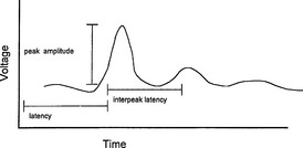
6 Describe the characteristics of the somatosensory-evoked potential waveform
The SSEP is plotted as a waveform of voltage vs. time. It is characterized by:


A waveform is identified by the letter describing its deflection above or below the baseline followed by a number indicating its latency (e.g., N20) (Figure 72-1).
7 Name several characteristic peaks important for the evaluation of somatosensory-evoked potentials
See Tables 72-1 and 72-2.
TABLE 72-1 Characteristic Peaks for Evaluation of Median Nerve Stimulation
| Peak | Generator |
|---|---|
| N9 | Brachial plexus (Erb’s point) |
| N11 | Dorsal root entry zone (cervical spine) |
| N13, 14 | Posterior column (nucleus cuneatus) |
| P14 | Medial lemniscus |
| N20 | Somatosensory cortex |
TABLE 72-2 Characteristic Peaks for Evaluation of Posterior Tibial Nerve Stimulation
| Peak | Generator |
|---|---|
| N20 | Dorsal root entry zone (lumbar spine) |
| N40 | Somatosensory cortex |
9 What are the indications for intraoperative use of somatosensory-evoked potential monitoring?


11 Summarize the effects of anesthetic agents on the amplitude and latency of somatosensory-evoked potentials
TABLE 72-3 Effects of Anesthetic Agents on Amplitude and Latency of Somatosensory-Evoked Potentials
| Drug | Amplitude | Latency |
|---|---|---|
| Premedication | ||
| Midazolam (0.3 mg/kg) | ↓ | 0 |
| Diazepam (0.1 mg/kg) | ↓ | ↑ |
| Induction agents | ||
| Thiopental (5 mg/kg) | ↑/0 | ↑ |
| Etomidate (0.4 mg/kg) | ↑↑↑ | ↑ |
| Propofol (0.5 mg/kg) | 0 | ↑ |
| Ketamine (1 mg/kg) | ↑ | * |
| Opioids | ||
| Fentanyl | ↑ | |
| Sufentanil | ↑ | |
| Morphine | ↑ | |
| Meperidine | ↑/↓ | ↑ |
| Inhaled anesthetics | ||
| Nitrous oxide | ↓ | ↑ |
| Isoflurane | ↓ | ↑ |
| Halothane | ↓ | ↑ |
| Enflurane | ↓ | ↑ |
| Desflurane | ↓ | ↑ |
| Sevoflurane | ↓ | ↑ |
| Others | ||
| Droperidol | ↓ | ↑ |
| Muscle relaxants | 0 | 0 |
↑, increase; ↓, decrease; 0, no change; *, not known.
12 What is the take-home message of the effects of anesthetic agents on somatosensory-evoked potentials?
 All of the halogenated inhaled anesthetics probably cause roughly equivalent dose-dependent decreases in amplitude and increases in latency that are further worsened by the addition of 60% nitrous oxide. It is best to restrict the use of volatile anesthetics and nitrous oxide to levels below 1 minimum alveolar concentration (MAC) and not to combine the two.
13 What other physiologic variables can alter somatosensory-evoked potentials?
 Temperature: Hypothermia increases latency, whereas amplitude is either decreased or unchanged. For each decrease of 1° C, latency is increased by 1 ms. Hyperthermia (4° C) decreases amplitude to 15% of the normothermic value.


14 If somatosensory-evoked potentials change significantly, what can the anesthesiologist and surgeon do to decrease the insult to the monitored nerves?


1. Jameson L.C., Janik D.J., Sloan T.B. Electrophysiologic monitoring in neurosurgery. Anesthesiol Clin. 2007;25:605-630.
2. Jameson L.C., Sloan T.B. Monitoring of the brain and spinal cord. Anesthesiol Clin. 2006;24:777-791.
3. Mahla M.E., Black S., Cucchiara R.F. Neurologic monitoring. In Miller R., editor: Anesthesia, ed 6, Philadelphia: Churchill Livingstone, 2005.
