CASE 72
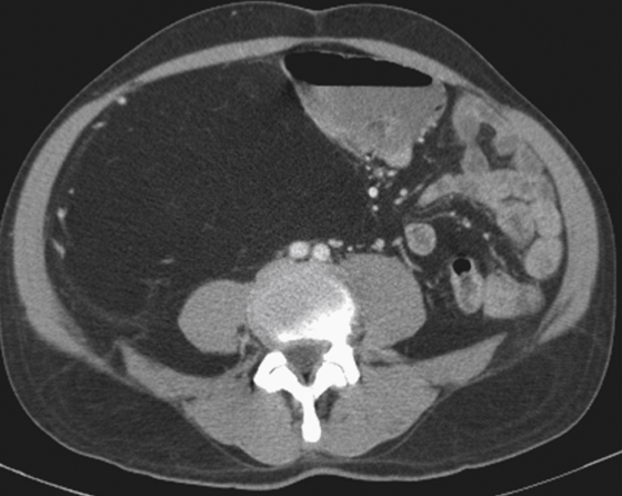
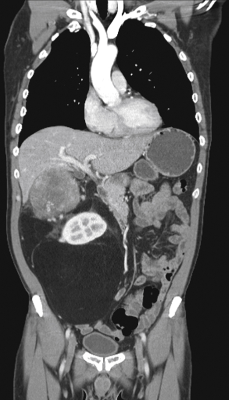
History: A 42-year-old man presents with diffuse, gradual abdominal enlargement and abdominal pain.
1. Which of the following should be included in the differential diagnosis of the imaging finding shown? (Choose all that apply.)
2. Which of the following statements regarding liposarcoma is true?
A. Liposarcoma typically occurs in children.
B. Liposarcoma usually arises from a pre-existing lipoma.
C. Liposarcoma is appropriately treated with radiotherapy.
D. Liposarcoma is encapsulated and therefore easily resected.
3. In adults, what is the most common retroperitoneal soft tissue sarcoma?
D. Malignant fibrous histiocytoma
4. Which of the following statements relevant to imaging of liposarcomas is true?
A. The most common location is the retroperitoneum.
B. A retroperitoneal fatty tumor is most likely a liposarcoma.
C. Liposarcoma usually invades the adjacent kidney and ureter.
D. The most common intra-abdominal location is the anterior perirenal space.
ANSWERS
CASE 72
Liposarcoma of the Abdomen
1. A, C, D, and E
2. C
3. A
4. B
References
Kim T, Murakami T, Oi H, et al: CT and MR imaging of abdominal liposarcoma. Am J Roentgenol. 1996;166:829–833.
Cross-Reference
Gastrointestinal Imaging: THE REQUISITES, 3rd ed, p 279.
Comment
Liposarcomas are relatively uncommon lesions seen in approximately 5000 patients in the United States annually. They are lipogenic tumors that can become widespread in the abdomen and extremely difficult to remove surgically. They are seen more commonly in adults, as opposed to the small, well-contained lipomas of the gut seen in children and young adults. Liposarcomas are rare in children. They can be mostly lipomatous with some soft tissue component, such as in this case, or almost purely lipomatous. They occur slightly more in male patients, and the less well differentiated the lesion, the poorer the outlook. Most patients have few or no symptoms until the tumor reaches a very large size. Eventually the patients become aware of swelling or pain and consult a physician. The imaging modality of choice is CT, in which the size, nature, and extent of involvement can be well described. CT diagnosis is usually quite accurate (see figures). Plain film can be helpful in determining the presence of a mass.


