CASE 72
1. What should be included in the differential diagnosis? (Choose all that apply.)
B. Thrombus
D. Aneurysm
2. What is a major advantage of CT angiography over nuclear medicine scintigraphic evaluation of chest pain?
A. Diagnosis of other diseases
D. Ability to evaluate sedentary patients
3. In addition to coronary artery stenosis involving the LAD coronary artery, what is the additional finding?
B. Thrombus
4. What is the recommendation for this patient?
ANSWERS
CASE 72
References
Kröner ES, van Velzen JE, Boogers MJ, et al. Positive remodeling on coronary computed tomography as a marker for plaque vulnerability on virtual histology intravascular ultrasound. Am J Cardiol. 2011;107(12):1725–1729.
Schoenhagen P, Ziada KM, Vince DG, et al. Arterial remodeling and coronary artery disease: the concept of “dilated” versus “obstructive” coronary atherosclerosis. J Am Coll Cardiol. 2001;38(2):297–306.
Zimmet JM, Miller JM. Coronary artery CTA: imaging of atherosclerosis in the coronary arteries and reporting of coronary artery CTA findings. Tech Vasc Interv Radiol. 2006;9(4):218–226.
Cross-Reference
Cardiac Imaging: The REQUISITES, ed 3, pp 248–261.
Comment
Imaging
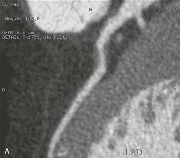
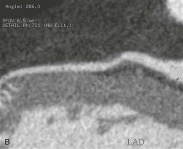
Multiplanar reformatted images from CT angiography show focal stenosis (<50%) of the LAD coronary artery by noncalcified plaque (Figs. A and B). The vessel is expanded outward in the region of atherosclerosis, which is known as positive remodeling.
Prognosis
The traditional thinking regarding coronary atherosclerosis is that it leads to progressive vessel narrowing as plaque is deposited within the intima; this is known as negative remodeling. However, certain plaque does not initially cause vessel narrowing but instead expands the media and external elastic membrane. This expansion causes the vessel to bulge outward in the region of atherosclerosis as a protective mechanism to limit the degree of luminal narrowing (Figs. A and B). This phenomenon is known as positive remodeling. In this process, the luminal cross-sectional area is not affected until the plaque causes a 40% area reduction. Catheter angiography is unable to detect the process of positive remodeling, whereas CT angiography can depict not only the lumen but also the outer wall. The process of positive remodeling is associated with vulnerable plaque owing to a larger lipid core and macrophage count; this helps explain why plaque rupture often occurs at sites of only minimal or modest stenosis. Negative remodeling is often associated with stable coronary syndromes. In the future, CT angiography may be used to identify positive remodeling because it is a marker of plaque vulnerability.


