CASE 71
History: No patient history is available.
1. In which patients is CT angiography of the coronary arteries indicated? (Choose all that apply.)
A. Patients with low pretest probability presenting with chest pain
B. Patients with intermediate pretest probability presenting with chest pain
E. Patients with chest pain and chest radiograph showing a pneumothorax
2. What percentage of patients presenting to the emergency department with chest pain are determined to have a coronary event at the time of discharge?
A. 5%
B. 25%
C. 50%
D. 80%
3. What reduces the specificity of CT angiography in accurately depicting the degree of coronary artery stenosis?
A. Coronary calcium score greater than 400
C. Extensive noncalcified plaque
4. What is the recommendation for this patient?
ANSWERS
Reference
Zimmet JM, Miller JM. Coronary artery CTA: imaging of atherosclerosis in the coronary arteries and reporting of coronary artery CTA findings. Tech Vasc Interv Radiol. 2006;9(4):218–226.
Cross-Reference
Cardiac Imaging: The REQUISITES, ed 3, pp 248–261.
Comment
Imaging
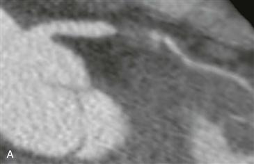
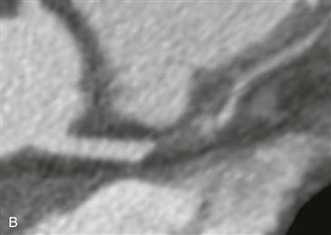
Two multiplanar reformatted images from CT angiography show complete occlusion of the left anterior descending (LAD) coronary artery by noncalcified plaque (Figs. A and B). The distal LAD is diminutive. LAD occlusion was confirmed via cardiac catheterization with distal reconstitution of the LAD via collaterals. Percutaneous stent placement was performed after determining the presence of residual myocardial viability with delayed thallium imaging.
Management
Coronary angiography remains the gold standard to assess coronary anatomy and to determine the degree of vessel narrowing. The main limitation of the test is that it is an invasive procedure with potential serious complications. CT angiography is useful in the evaluation of patients with a low or intermediate pretest probability of disease. A negative test essentially rules out significant pathology. The patient is able to be discharged earlier, and there is evidence of significant cost savings. A positive test must be confirmed with invasive coronary angiography. In this case, the patient has a positive CT angiogram with complete LAD occlusion and requires confirmation and potentially treatment with coronary angiography.


