[level-membership-for-anesthesiology-category]
CHAPTER 7 Coagulation
5 What are the intrinsic and extrinsic coagulation pathways?
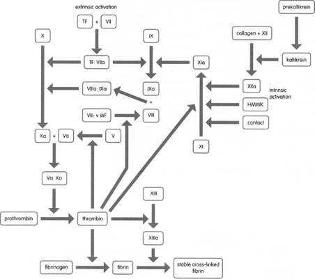
Traditionally these two pathways have been viewed as separate mechanisms that merge after the formation of activated factor X (Figure 7-1). This rigid division has lost absolute validity because of the crossover of many factors. For instance, factor VIIa can activate factor IX; but factors IXa, Xa, thrombin, and XIIa can activate factor VII. However the classic two-pathway model is still useful for the interpretation of in vitro coagulation studies.


11 Review the properties of factor VIII

22 What is disseminated intravascular coagulation?


27 Discuss the parameters measured by thromboelastography
There are five parameters of the TEG tracing: R, k, alpha angle, MA, and MA60 (Figure 7-2).


 MA60: Measures the rate of amplitude reduction 60 minutes after MA, representing the stability of the clot (Figure 7-2)
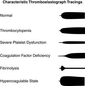

1. Drummond J.C., Petrovitch C.T. Hemostasis and hemotherapy. In: Barash P.G., Cullen B.F., Stoelting R.K., editors. Clinical anesthesia. ed 5. Philadelphia: Lippincott, Williams & Wilkins; 2006:221-240.
2. Wenker O., et al. Thrombelastography. The Internet Journal of Anesthesiology. http://www.ispub.com.
[/level-membership-for-anesthesiology-category][not-level-membership-for-anesthesiology-category]
CHAPTER 7 Coagulation
5 What are the intrinsic and extrinsic coagulation pathways?
Traditionally these two pathways have been viewed as separate mechanisms that merge after the formation of activated factor X (Figure 7-1). This rigid division has lost absolute validity because of the crossover of many factors. For instance, factor VIIa can activate factor IX; but factors IXa, Xa, thrombin, and XIIa can activate factor VII. However the classic two-pathway model is still useful for the interpretation of in vitro coagulation studies.
