CASE 7
1. What should be included in the differential diagnosis for Fig. A? (Choose all that apply.)
A. Fat pad
D. Esophageal duplication cyst
2. Where does this lesion most commonly occur?
3. What is the most likely internal composition of this lesion?
A. Fat
B. Soft tissue
C. Fluid
D. Calcium
4. What is the next step in management in this asymptomatic patient?
A. Nothing
B. Aspiration
ANSWERS
Reference
Wang ZJ, Reddy GP, Gotway MB, et al. CT and MR imaging of pericardial disease. Radiographics 2003;.
Cross-Reference
Cardiac Imaging: The REQUISITES, ed 3, pp 56, 78–80, 139–141, 277.
Comment
Epidemiology
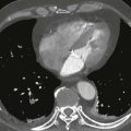
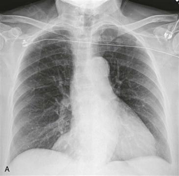
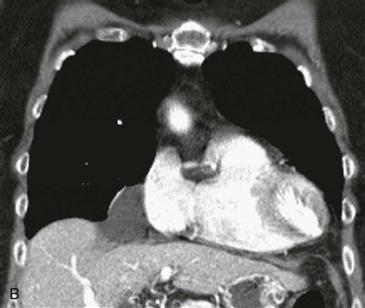
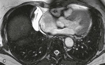
Pericardial cysts are congenital lesions formed when pericardium is pinched off during development. Most pericardial cysts are located in the right cardiophrenic angle followed by the left cardiophrenic angle. Cysts can be readily distinguished from other cardiophrenic angle masses with cross-sectional imaging (either CT or MRI). Pericardial cysts are well-defined, nonenhancing lesions of fluid attenuation. They are uniformly hyperintense on T2-weighted MRI.
Presentation
Patients with pericardial cysts are usually asymptomatic, but large cysts can cause dyspnea, chest pain, cough, and rarely cardiac tamponade. Treatment of symptomatic lesions is surgical resection or percutaneous drainage.