CASE 68

History: A 78-year-old woman presents feeling very unwell and with abdominal pain.
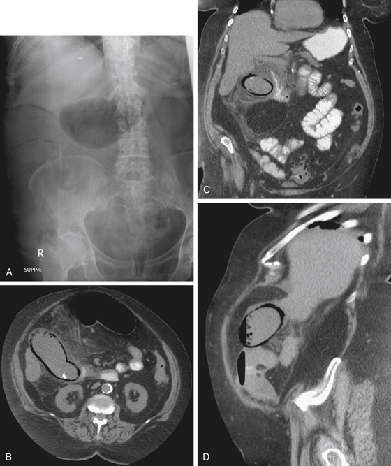
1. Which of the following should be included in the differential diagnosis of the imaging finding shown in Figure A? (Choose all that apply.)
D. Emphysematous cholecystitis
E. Emphysematous pyelonephritis
2. What organism typically produces the process shown in these images of the right upper quadrant?
3. What underlying condition does this patient probably have?
4. What is the major complication leading to a high mortality rate?
D. Common bile duct obstruction
ANSWERS
CASE 68
Emphysematous Cholecystitis
1. A, B, C, and D
2. D
3. D
4. A
References
Gill KS, Chapman AH, Weston MJ. The changing face of emphysematous cholecystitis. Br J Radiol. 1997;70:986–991.
Cross-Reference
Gastrointestinal Imaging: THE REQUISITES, 3rd ed, p 255.
Comment
The appearance of gas in the lumen or the wall of the gallbladder is diagnostic of a condition known as emphysematous cholecystitis. The gas is the result of the gas-forming organism that is causing the cholecystitis. Most commonly, clostridial organisms are to blame. Patients who develop this condition are typically diabetic; rarely does emphysematous cholecystitis occur in persons without diabetes as an underlying condition. Often the patient is older and has had diabetes for many years, resulting in vascular insufficiency to the gallbladder, which is probably a major underlying cause for the development of emphysematous cholecystitis. Usually, cystic duct obstruction is also present. Although gas is present in the gallbladder lumen, gas is rarely encountered in the rest of the biliary system. The risk of perforation in patients with emphysematous cholecystitis is five times higher than that in patients with ordinary acute cholecystitis.
Emphysematous cholecystitis is one of the few diagnoses that can be readily made based on conventional radiographs (see figures). When the patient is experiencing severe abdominal pain and there is gas in the gallbladder lumen, particularly if the patient is diabetic, the diagnosis of emphysematous cholecystitis must be a primary consideration. Many conditions can result in the accumulation of gas in the lumen of the gallbladder, but there is usually an abnormal connection of the biliary tract to the bowel lumen allowing this to occur. Also, in some instances gas is visible in the wall of the gallbladder, indicating necrosis of the gallbladder wall. On CT the abnormalities are shown quite well and pericholecystic complications of inflammation and abscess are demonstrated (see figures). This condition might not be readily diagnosed on ultrasound, however, because the gas can mimic gallstones and produce acoustic shadowing.


