CASE 68
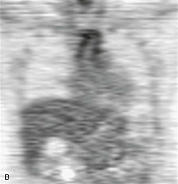
1. What should be included in the differential diagnosis for fluorodeoxyglucose (FDG) uptake shown in Fig. B? (Choose all that apply.)
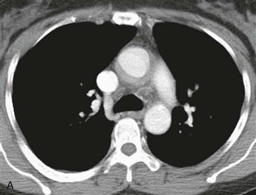
2. What is the imaging finding seen in Fig. A?
D. Intimal flap
3. What is the most likely diagnosis in this 35-year-old woman presenting with arm claudication and an elevated sedimentation rate?
4. What is the role of PET in this disease?
A. Monitor response to therapy
B. No role
ANSWERS
Reference
James OG, Christensen JD, Wong TZ, et al. Utility of FDG PET/CT in inflammatory cardiovascular disease. Radiographics. 2011;31(5):1271–1286.
Cross-Reference
Cardiac Imaging: The REQUISITES, ed 3, pp 393–394.
Comment
Differential Diagnosis
Axial contrast-enhanced CT shows ascending aortic wall thickening with enhancement (Fig. A). Coronal PET shows increased FDG activity in the ascending aortic wall (Fig. B). The main differential diagnoses include atherosclerosis and a large vessel vasculitis. The two main large vessel vasculitides are Takayasu arteritis and giant cell arteritis. Takayasu arteritis most commonly affects females 10 to 40 years old, whereas giant cell arteritis occurs in patients older than 50 years.
Monitoring Response to Therapy
The role of PET in diagnosing and monitoring treatment response in patients with an inflammatory arteritis is evolving. Often PET may be the first study to suggest a vasculitis when it is performed in patients referred for fever of unknown origin or nonspecific constitutional symptoms. The characteristic finding of a large vessel vasculitis is circumferential increased FDG activity within the involved vessel wall (Fig. B). This increased FDG activity represents active disease and is relatively sensitive and specific for the diagnosis in the presence of elevated inflammatory markers. PET is also useful in monitoring disease response to antiinflammatory medications. PET shows treatment response earlier than anatomic imaging. Decreased vessel wall metabolic activity correlates with symptom resolution and reduced inflammatory blood markers.


