[level-membership-for-gastroenterology-and-hepatology-category]CASE 66
History: A 68-year-old woman presents with abnormal liver function tests.
1. Which of the following should be included in the differential diagnosis of the imaging finding shown in the figures? (Choose all that apply.)
2. Histologically, of what tissue type are most duodenal polyps?
3. What is the most common cause of polypoid-like filling defects at the base of the duodenal bulb?
B. Brunner’s gland hyperplasia
4. With which polyposis syndrome is small bowel and duodenal polyps most commonly associated?
ANSWERS
CASE 66
Villous Adenomas of the Duodenum
1. A, B, C, and E
2. A
3. C
4. B
References
Dahnert W. Radiology Review Manual. 6th ed Philadelphia: Lippincott Williams & Wilkins; 2007. p 774
Cross-Reference
Gastrointestinal Imaging: THE REQUISITES, 3rd ed, p 94.
Comment
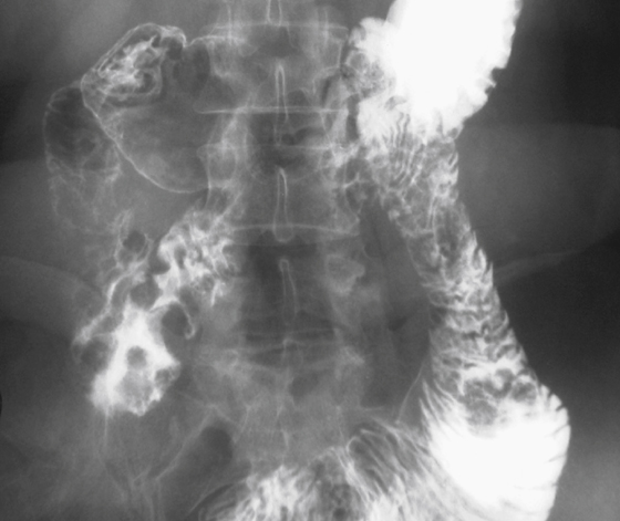
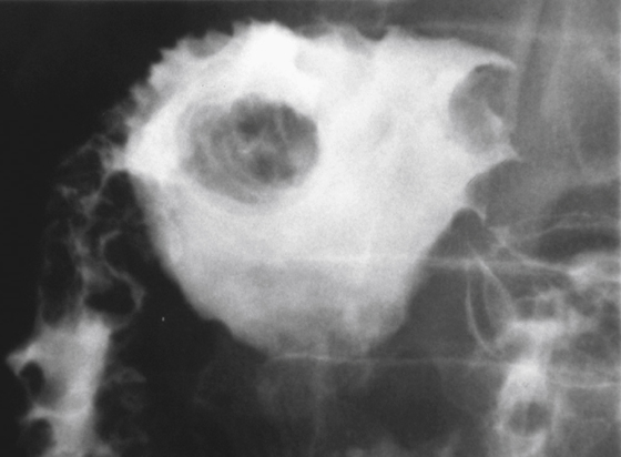
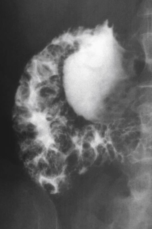
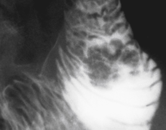
Once thought to be rare, in recent decades the occurrence of solitary polyps in the duodenum has been found to be more common than once thought. In contrast to the stomach, where most polyps are hyperplastic, in the duodenum, solitary polyps are most often adenomas and dangerous. Hyperplastic polyps of the duodenum are rare, despite the inflammatory changes that occur there. As is true of colonic polyps, the larger the adenoma, the more likely it is to be malignant. However, unlike the colon, any adenomatous polyp in the duodenum has a greater risk for malignancy.
The location of the polyp may be a helpful distinguishing feature. A polypoid filling defect in the bulb may be a gastric polyp, prolapsing through the pylorus along with gastric mucosa, a Brunner’s gland adenoma (not a true adenomatous polyp), another type of tumor (e.g., gastrointestinal stromal tumor [GIST], metastasis), an adenoma, or an ectopic pancreatic rest. The more distal the polyp is in the duodenum, the more likely it is to be an adenoma or villous adenoma. Villous adenomas are particularly common in the periampullary region and can result in biliary and pancreatic duct obstruction. GIST tumors and ectopic pancreatic rests can occur anywhere but are more common in the proximal half of the duodenum. As a general rule, the more distal the lesion is in the duodenum, the more likely it is to be clinically important.
Duodenal polyps are more common in the patient with virtually any of the polyposis syndromes (see figures). In the patient with familial polyposis coli, the growths may be either adenomas or hyperplastic polyps. Patients with Gardner’s syndrome or familial polyposis coli have a high incidence of periampullary malignancies, particularly those with Gardner’s syndrome.
[/level-membership-for-gastroenterology-and-hepatology-category][not-level-membership-for-gastroenterology-and-hepatology-category]CASE 66
History: A 68-year-old woman presents with abnormal liver function tests.
1. Which of the following should be included in the differential diagnosis of the imaging finding shown in the figures? (Choose all that apply.)
2. Histologically, of what tissue type are most duodenal polyps?
3. What is the most common cause of polypoid-like filling defects at the base of the duodenal bulb?
[/not-level-membership-for-gastroenterology-and-hepatology-category]
