CASE 65
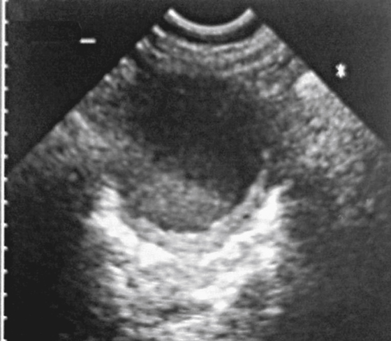
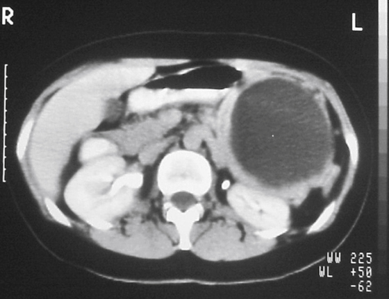
History: A 39-year-old woman presents with 3 months of pain in the left hypochondrium. On examination, a mass is easily palpable in the left upper quadrant.
1. Which of the following should be included in the differential diagnosis of the imaging finding shown in the figures? (Choose all that apply.)
2. Which statement concerning mucinous cystadenoma of the pancreas is true?
A. They most often occur in the pancreatic head.
B. They have no malignant potential.
C. Calcification is very rare.
D. Average age of patients is 50 years.
E. Typically there are multiple cysts.
3. Which of the following statements regarding pancreatic microcystic adenomas is true?
A. This tumor is similar on imaging to mucinous tumors.
B. On CT, the solid component of the tumor is nonenhancing.
C. Calcification contributes to a well-defined surrounding capsule.
D. The tumor is associated with von Hippel–Lindau disease.
4. Which is the most common of these pancreatic tumors?
ANSWERS
CASE 65
Cystic Tumor of the Pancreas
1. A, B, C, and E
2. D
3. D
4. C
References
Lundstedt C, Dawiskiba S. Serous and mucinous cystadenoma/cystadenocarcinoma of the pancreas. Abdom Imaging. 2000;25:201–206.
Cross-Reference
Gastrointestinal Imaging: THE REQUISITES, 3rd ed, p 166.
Comment
Several neoplasms of the pancreas manifest as cystic lesions, although all are fairly rare compared with ductal adenocarcinoma, which almost never has a cystic component. Central necrosis is usually easily distinguishable from a cystic component.
The term mucinous cystic neoplasms describes what has been termed mucinous cystadenoma and mucinous cystadenocarcinoma. These tumors are difficult to distinguish, and because cystadenomas have malignant potential, they are now classified together and all are considered malignant or potentially malignant. Often the cyst is unilocular, resembling a pseudocyst, or cysts can be large, with multiple distinct septations. These tumors are hypovascular, and if they calcify, they do so in a peripheral location.
Another cystic tumor is microcystic adenoma, which has multiple small cysts of varying size. If the cysts are too numerous to count, the diagnosis should be considered microcystic adenoma. This condition occurs in older patients, mostly female. This tumor is associated with von Hippel–Lindau disease. It is a hypervascular tumor that develops central necrosis and scarring, often with calcification.
Another tumor that can resemble a mucinous cystic neoplasm is a cystic teratoma, although this tumor is rare. This tumor also has multiple large cysts and dystrophic calcification. Papillary epithelial neoplasms can be cystic. The cyst is usually thick walled, with mural tumor projections. This tumor occurs in young women and is considered a low-grade malignancy. Also, islet cell tumors that undergo necrosis can show cystic changes.


