CASE 64
1. What should be included in the differential diagnosis for a left-to-right shunt? (Choose all that apply.)
B. Partial anomalous pulmonary venous return (PAPVR)
C. Ventricular septal defect (VSD)
D. Patent ductus arteriosus (PDA)
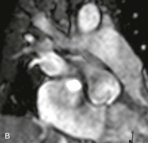
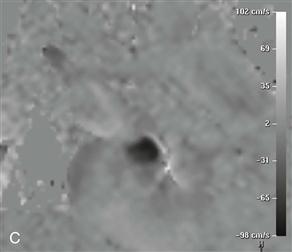
2. What imaging sequence is shown in Figs. B and C?
A. Steady-state free precession (SSFP)
B. Velocity-encoded cine (VEC) phase contrast
D. Late gadolinium enhancement
3. What is the most debilitating complication of this entity?
A. Paradoxical embolism with stroke
4. What is the most likely diagnosis?
A. Cardiac mass
C. VSD
ANSWERS
Reference
Dodd JD, Aquino SL, Holmvang G, et al. Cardiac septal aneurysm mimicking pseudomass: appearance on ECG-gated cardiac MRI and MDCT. AJR Am J Roentgenol. 2007;188(6):W550–W553.
Comment
Diagnosis
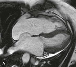
Atrial septal aneurysm is uncommon; it is a discrete bulge of the septum into either the left or the right atrium (Fig. A). To be considered an aneurysm, it must protrude greater than 1.0 to 1.5 cm beyond the expected plane of the atrial septum, and the aneurysm neck must measure more than 1.5 cm in diameter. Thrombus may form within the aneurysm; given the high rate of patent foramen ovale and ASD, there is increased risk of paradoxical embolism and stroke. Care must be taken not to confuse this entity with a cardiac mass on routine nongated chest CT because there may be unopacified blood owing to lack of mixing of contrast material.
Cardiac MRI
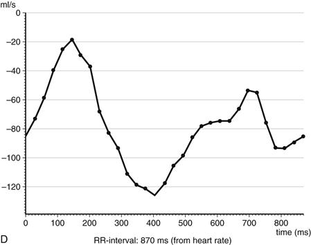
Cardiac MRI has the added value of quantifying the shunt fraction through the use of VEC phase contrast MRI (Figs. B–D). As shown in this case, the shunt can be directly quantified with phase imaging by choosing a plane perpendicular to the aneurysm. An additional method of quantifying the left-to-right shunt in the absence of other valvular abnormalities would be to calculate the pulmonic-to-systemic flow (Qp/Qs) ratio with VEC phase contrast MRI. The Qp/Qs ratio is calculated by performing phase images directly above the pulmonic and aortic valves. In normal patients, the blood leaving the left and right ventricles should be roughly equal. In patients with a shunt, the Qp/Qs ratio does not equal 1 and is directly related to the shunted fraction.


