History: A patient without relevant symptoms, with an incidental abnormality noted on chest x-ray, was referred for further imaging.
1. Which of the following should be included in the differential diagnosis of the imaging findings shown for this patient? (Choose all that apply.)
2. All of the following are foregut congenital cysts except:
3. What is the most common symptom of an esophageal duplication cyst?
4. Which of the following statements about imaging of esophageal duplication cysts is true?
A. Radionuclide scanning with 99mTc pertechnetate is positive in half of the cases.
B. On barium swallow, the cyst typically appears as an intramural diverticulum.
D. On MRI, the lesion typically has short T1 and long T2 relaxation times.
ANSWERS
CASE 6
Duplication Cyst of the Esophagus
1. A, C, and D
2. C
3. A
4. A
Reference
Jeung MY, Gasser B, Gangi A, et al: Imaging of cystic masses of the mediastinum. Radiographics. 2002;22:S79–S93.
Cross-Reference
Gastrointestinal Imaging: THE REQUISITES, 3rd ed, p 10.
Comment
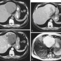
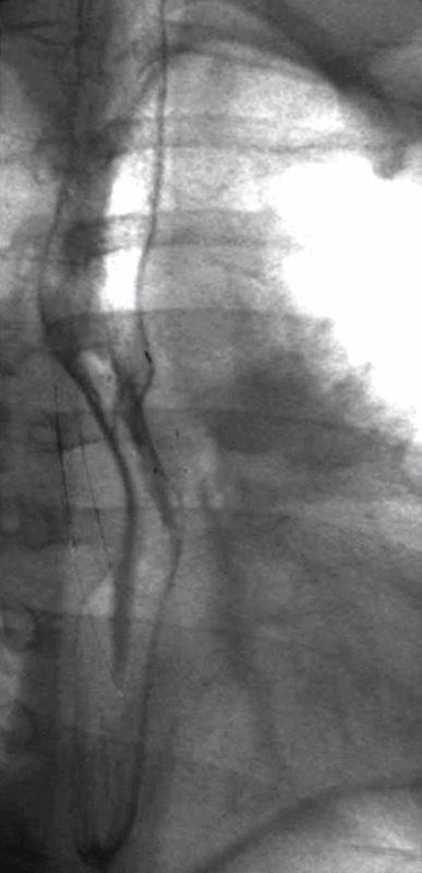
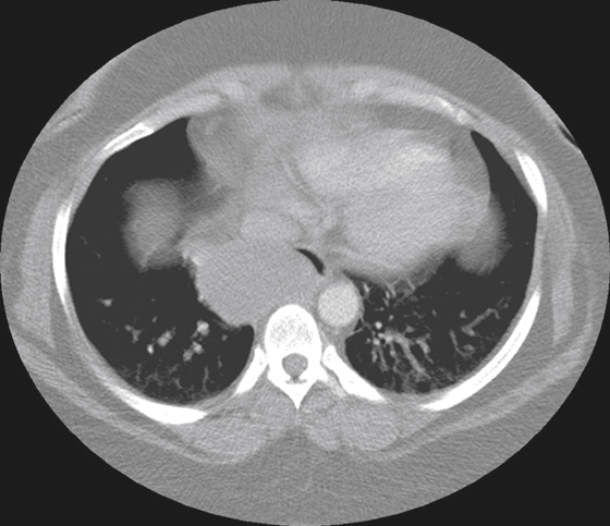
The barium swallow discloses an extrinsic impression on the lower esophagus (see figures). The angles of the impression are obtuse, the mucosa is smooth and intact, and the lumen is somewhat compromised. There is no evidence of ulceration or mucosal destruction. All these findings suggest an extrinsic origin. In fact, the lesion is a noncommunicating duplication cyst of the esophagus. A small percentage of cysts communicate with the esophagus and can act like an epiphrenic diverticulum if the cyst is located in the lower esophagus. Esophageal duplication cysts of the gut are the most common enteric cyst (25%). Duplication cysts are one of the cystic manifestations of foregut malformations.
CT demonstrates a paraesophageal cystic mass, which usually is not communicating with the lumen (see figures). A small percentage communicate. Bronchogenic and neurogenic cysts are the other two foregut malformation cysts, and all can affect the esophagus. These cysts tend to be unilocular and bear a close embryologic relationship to congenital cystic adenomatoid malformation of the lung. Duplication cysts are usually lined with squamoid cells with some columnar elements and are usually seen in the lower esophagus.
Bronchogenic cysts are usually indistinguishable and are most commonly seen in the mediastinum in a subcarinal location. They usually contain some ciliated columnar epithelium and are thought to be the result of abnormal bronchial budding during lung development. They never communicate with the esophagus. Neurogenic cysts are usually located in the posterior mediastinum and in a more posterior position than either duplication or bronchogenic cysts. They are thought to be an embryologic abnormality associated with incomplete separation from the notochordal structures. Neurogenic cysts are commonly associated with vertebral abnormalities such as hemivertebrae or butterfly vertebrae.