[level-membership-for-cardiovascular-category]
CASE 6
1. What should be included in the differential diagnosis for these two patients? (Choose all that apply.)
C. Hypertrophic cardiomyopathy
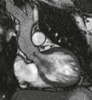
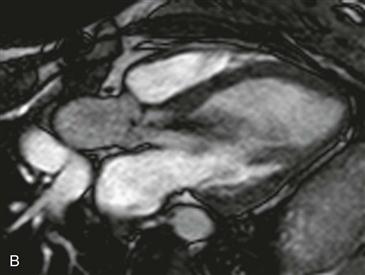
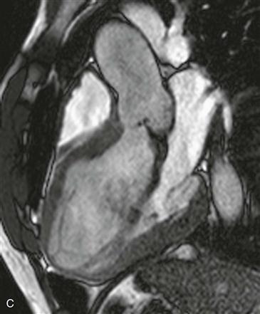
2. What physical fluid property is responsible for the signal void (black signal) that is seen directed into the left ventricle from the aortic valve?
A. Steady flow
B. Laminar flow
3. What is the most frequent cause of the finding in Figs. A–C?
B. Idiopathic valvular degeneration
D. Endocarditis
4. Which imaging sequence could quantify the abnormality shown in Figs. A–C?
ANSWERS
Reference
Glockner JF, Johnston DL, McGee KP. Evaluation of cardiac valvular disease with MR imaging: qualitative and quantitative techniques. Radiographics. 2003;23(1):e9.
Cross-Reference
Cardiac Imaging: The REQUISITES, ed 3, pp 182–185.
Comment
Epidemiology
Aortic regurgitation most commonly occurs in elderly patients with senile degeneration of the aortic valve and is often seen in association with aortic stenosis. In patients younger than 40 years of age, aortic regurgitation is often due to Marfan syndrome with aortic root dilation. MRI is ideally suited not only to quantify the volume of regurgitation but also to determine its overall effect on the heart. Velocity-encoded cine phase contrast MRI accurately and reproducibly quantifies the volume of valve regurgitation through measurement of the regurgitant fraction (regurgitant volume/forward stroke volume). Cine SSFP images are able to evaluate the overall effect of the regurgitant valve on the heart through measurement of chamber size, ejection fraction, and cardiac mass. Some authors advocate valve replacement in asymptomatic patients because irreversible damage and ventricular remodeling may have already occurred if the valve is replaced after onset of symptoms.
[/level-membership-for-cardiovascular-category][not-level-membership-for-cardiovascular-category]
CASE 6
1. What should be included in the differential diagnosis for these two patients? (Choose all that apply.)
C. Hypertrophic cardiomyopathy
2. What physical fluid property is responsible for the signal void (black signal) that is seen directed into the left ventricle from the aortic valve?
A. Steady flow
B. Laminar flow
[/not-level-membership-for-cardiovascular-category]
