CASE 59
1. What should be included in the differential diagnosis? (Choose all that apply.)
A. Cyst
B. Lipoma
C. Metastasis
D. Hematoma
2. What is the most likely location of the mass?
A. Pericardium
B. Myocardium
C. Left atrium
D. Right atrium
3. What is the most likely diagnosis?
A. Cyst
B. Lipoma
C. Metastasis
D. Hematoma
4. What is the most appropriate management?
A. No treatment
B. Chemotherapy
D. Surgery
ANSWERS
Reference
Yared K, Baggish AL, Picard MH, et al. Multimodality imaging of pericardial diseases. JACC Cardiovasc Imaging. 2010;3(6):650–660.
Cross-Reference
Cardiac Imaging: The REQUISITES, ed 3, p 273.
Comment
Clinical Features
Cardiac lipoma most commonly occurs in the right atrium. It is the second most common benign primary cardiac neoplasm following myxoma. Other locations include the epicardium, endocardium, interatrial septum, and left ventricle. Pericardial lipomas are less common, but they are among the more common masses of the pericardium. Lipomas tend to be soft and flexible, and even large tumors may not compress the heart. They are usually asymptomatic. If there are symptoms of compression, such as pain or shortness of breath, then surgical resection is usually performed.
CT and MRI Features
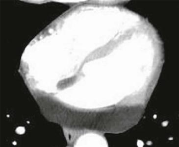
On CT scan, a pericardial lipoma manifests as a homogeneous, low-density mass (negative Hounsfield units) in the pericardium that is relatively pliable without much mass effect (Figure). There is generally no enhancement and no signs of invasion. Fat saturation MRI sequences are ideal for demonstrating that the mass is composed of fat.


