CASE 57
1. What should be included in the differential diagnosis? (Choose all that apply.)
D. Lymphoma
2. Which structure is most severely compressed?
B. Aorta
C. Bronchus
3. What is the most likely diagnosis?
D. Hematoma
4. What is the most likely location of the mass?
C. Myocardium
D. Pericardium
ANSWERS
Reference
Yared K, Baggish AL, Picard MH, et al. Multimodality imaging of pericardial diseases. JACC Cardiovasc Imaging. 2010;3(6):650–660.
Cross-Reference
Cardiac Imaging: The REQUISITES, ed 3, pp 102–104.
Comment
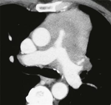
Radiographic Features
Most pericardial tumors are secondary to direct extension or hematogenous spread from lung or breast carcinoma, lymphoma, and melanoma. Findings suggestive of pericardial malignancy include enhancing pericardial nodules or masses and hemopericardium. Primary pericardial tumors are rare; mesothelioma is the most common. Other primary pericardial tumors include lymphoma and teratoma.
Primary Cardiac Lymphoma
Primary pericardial lymphoma is associated with acquired immunodeficiency syndrome (AIDS) and is generally a non-Hodgkin lymphoma. By definition, the lymphoma is localized to the heart or pericardium at diagnosis without evidence of systemic disease. Presenting symptoms include rapidly progressive heart failure, arrhythmia, cardiac tamponade, superior vena cava syndrome, and chest pain. Pleural effusion, including hemorrhagic effusion, occurs frequently. The prognosis of primary cardiac lymphoma is poor with many patients being diagnosed at autopsy. Treatment includes chemotherapy and tumor debulking for palliative control of symptoms.
Imaging
CT is frequently performed for diagnosis, but MRI is especially useful to define the extent of involvement. Lymphoma is typically infiltrative and follows the pericardial contour (Figure). Both CT and MRI show heterogeneous enhancement following the administration of intravenous contrast. Cross-sectional imaging plays an important role in assessing disease response to therapy.


