CASE 56
History: A 28-year-old woman presents with dyspepsia and vomiting and a history of alcohol and ibuprofen abuse.
1. Which of the following should be included in the differential diagnosis of an underlying disease resulting in the imaging finding shown in the figures? (Choose all that apply.)
2. Which choice is the most likely diagnosis?
3. Which of the following are features of a benign gastric ulcer?
D. Ulcer in profile projects within the gastric lumen
E. Radiating folds extending to surrounding edema mound
4. Which of the following is the most common cause of gastric and duodenal peptic ulcer disease in industrialized societies?
C. Use of nonsteroidal antiinflammatory drugs (NSAIDs)
E. Helicobacter pylori infection
ANSWERS
CASE 56
Benign Gastric Ulcers
1. A, B, C, and E
2. B
3. A
4. E
References
Levine MS. Peptic ulcers. In: Gore RM, Levine MS, eds. Textbook of Gastrointestinal Radiology. 2nd ed Philadelphia: WB Saunders; 2000:514–545.
Cross-Reference
Gastrointestinal Imaging: THE REQUISITES, 3rd ed, p 82.
Comment
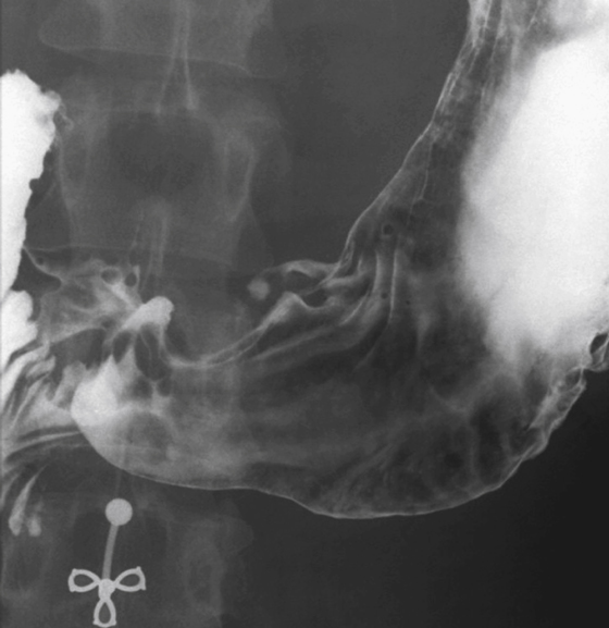
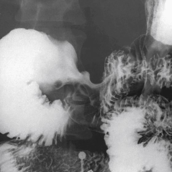
Benign ulcers (see figures) can occur anywhere in the stomach, although they do have a propensity to occur in the lesser curvature, particularly in the middle of the body. This type of ulcer is often seen in the older patient, but the cause for this is uncertain. As a corollary, the majority of ulcers found in the fundus are malignant. The size of an ulcer has no bearing on its malignant potential, and giant gastric ulcers often represent a penetrating, walled-off, benign gastric ulcer. Also, the shape of the ulcer is usually round and well defined, but this is not always the case.
Much has been said about the gastric ulcer as seen in profile. Most benign gastric ulcers project outside the expected lumen of the stomach. However, if there is a great deal of edema or the ulcer is chronic, this might not be the case. Another common sign of a benign gastric ulcer is the Hampton line sign, which is a thin lucent line at the neck or base of the ulcer as it passes through the mucosal layer where undermining of the submucosa occurs. However, a very thick ulcer collar, particularly if it is asymmetrical, at the neck of the ulcer is not a Hampton line and may be seen in benign as well as malignant ulcers.
Often, radiating folds are seen extending toward an ulcer. If these folds pass all the way to the edge of the crater, the process is most likely benign. Infrequently, folds stop short as a result of a large collar of edema (in benign ulcers) or as a result of neoplastic tissue (in malignant ulcers); therefore, folds that stop short are a good but not absolute differential diagnostic feature. Also, the folds tend to be smooth and symmetrical in benign ulcers and more irregular, thickened, and fused in malignant ulcers. It has long been a part of gastric ulcer lore that malignant ulcers can show signs of healing.


