CASE 56
History: A patient presents with chest pain.
1. What should be included in the differential diagnosis? (Choose all that apply.)
A. Cyst
B. Lipoma
C. Metastasis
D. Hematoma
2. What is the most likely location of the mass?
A. Pericardium
B. Myocardium
3. What is the most likely diagnosis?
A. Cyst
B. Lipoma
C. Metastasis
D. Hematoma
4. Which chamber is most severely compressed?
A. Left atrium
C. Right atrium
ANSWERS
Reference
Wang ZJ, Reddy GP, Gotway MB, et al. CT and MR imaging of pericardial disease. Radiographics. 2003;23(Spec No):S167–S180.
Cross-Reference
Cardiac Imaging: The REQUISITES, ed 3, pp 265, 270.
Comment
Etiology and Physiology
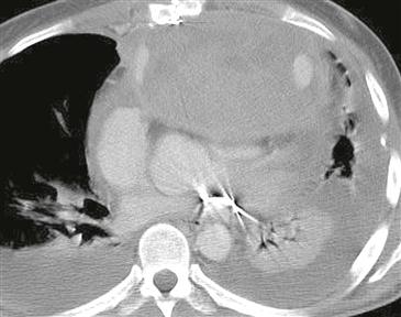
A pericardial hematoma can result from trauma (iatrogenic or otherwise), myocardial infarction, aortic dissection, tumor, or pericarditis. When a hematoma is large enough to compress a cardiac chamber and cause hemodynamic compromise, it may have to be evacuated to relieve the compression.
Differential Diagnosis
The differential diagnosis for a pericardial mass includes a pericardial cyst, tumor (metastatic or primary), and hematoma.
3. Pericardial hematoma is characterized by high or heterogeneous density on CT (Figure). A chronic hematoma can be calcified. MRI is useful as it may show characteristic high T1 and T2 signal from associated blood product degradation. Hematomas do not enhance following contrast administration.


