[level-membership-for-gastroenterology-and-hepatology-category]CASE 55
History: An 83-year-old man presents with lower limb pitting edema.
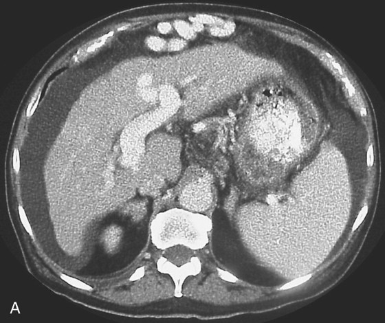
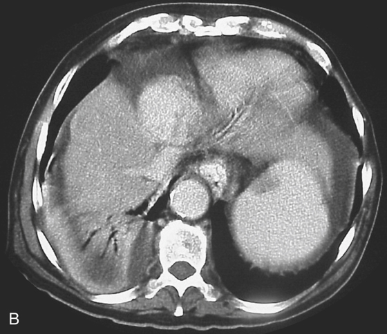
1. Which of the following should be included in the differential diagnosis of the imaging findings shown in figure A? (Choose all that apply.)
D. Chronic left atrial failure
E. Arteriovenous portal fistula
2. Name the leading cause for hepatic cirrhosis in North America.
D. Traumatic arteriovenous portal fistula
3. All of the following are some of the complications seen with portal hypertension except:
D. Right heart valvular fibrosis
4. With widespread cirrhotic involvement of the liver, what area may be spared or less affected?
ANSWERS
CASE 55
Portal Hypertension
1. A, B, and E
2. A
3. D
4. B
References
Baron RL, Gore RM. Diffuse liver disease. In: Gore RM, Levine MS, eds. Textbook of Gastrointestinal Radiology. 2nd ed Philadelphia: WB Saunders; 2000:1590–1638.
Cross-Reference
Gastrointestinal Imaging: THE REQUISITES, 3rd ed, p 184.
Comment
Cirrhosis of the liver is the result of chronic liver hepatocyte toxicity resulting in fibrous tissue formation throughout the liver, with resultant tissue distortion and a smaller shrunken liver. The changes may initially be focal. However, when the entire liver is involved, the classic CT appearance of a small liver with nodular or irregular margins is seen. The caudate lobe may be partially spared. Although most of the liver’s blood supply comes from the portal vein, the caudate lobe has a direct connection with the inferior vena cava in most patients. In the United States, most cirrhosis is alcohol induced (Laennec’s cirrhosis) and is seen with splenomegaly, ascites, and esophageal varices (see figures). The falciform ligament may also be seen if there is sufficient ascites (see figures).
Other causes of cirrhosis include Wilson’s disease and hemochromatosis. Portal vein hypertension is a form of relative occlusion of the portal vein. In some cases of cirrhosis a true portal vein thrombus may be seen. However, there also may be other causes of portal vein thrombosis apart from cirrhosis.
[/level-membership-for-gastroenterology-and-hepatology-category][not-level-membership-for-gastroenterology-and-hepatology-category]CASE 55
History: An 83-year-old man presents with lower limb pitting edema.
1. Which of the following should be included in the differential diagnosis of the imaging findings shown in figure A? (Choose all that apply.)
D. Chronic left atrial failure
E. Arteriovenous portal fistula
2. Name the leading cause for hepatic cirrhosis in North America.
D. Traumatic arteriovenous portal fistula
[/not-level-membership-for-gastroenterology-and-hepatology-category]
