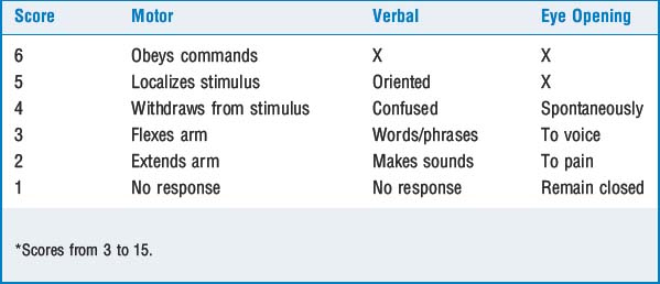
54: Trauma

Published on 06/02/2015 by admin

Filed under Anesthesiology

Last modified 22/04/2025

Print this page

rate 1 star rate 2 star rate 3 star rate 4 star rate 5 star
Your rating: none, Average: 0 (0 votes)

This article have been viewed 1889 times

CHAPTER 54 Trauma

7 How does an uncleared cervical spine modify the approach to the airway?

Patients requiring emergent surgical procedures do not have time to have their cervical spines evaluated fully. There is no airway management technique that results in no cervical motion. However, there is no documentation of iatrogenic neurologic injury in patients with cervical fractures when cervical spine precautions are used. These precautions include an appropriately sized Philadelphia collar, sand bags placed on each side of the head and neck, and the patient resting on a hard board with the forehead taped and secured to it.

Alternative airway management techniques in the traumatized patient include rapid-sequence induction with in-line stabilization, use of the Bullard laryngoscope, blind nasal intubation, and fiber-optic bronchoscopic-assisted ventilation. A Glidescope is a laryngscope with a camera lens on its tip and is very useful when a patient’s neck must be maintained in a neutral position. An unstable or uncooperative patient likely would receive a rapid-sequence induction.

When a cervical fracture or cervical spinal cord injury (SCI) is documented, most anesthesiologists choose fiber-optic intubation facilitated by some form of topical anesthesia to the airway and sedation, titrated to effect, keeping in mind the patient’s other injuries and hemodynamic status. This allows postintubation assessment of neurologic status before induction of unconsciousness. It would not be advisable to ablate all protective airway reflexes in a patient with a full stomach.

16 What challenges do spinal cord–injured patients pose?

Airway management has been discussed briefly. The technique of choice depends on the urgency of the situation but might be direct laryngoscopy with in-line stabilization, intubation using a Bullard laryngoscope, or fiberoptic intubation. When moved, patients should be log-rolled; that is, rolled and moved with care to maintain the neck in a neutral position.

Some degree of neurogenic shock should be expected with injuries above the T6 level. However, hypotension in cord-injured patients is most likely caused by other injures. Catecholamine surges may produce pulmonary vascular damage, resulting in neurogenic pulmonary edema. There may be some element of myocardial dysfunction. If moderate fluid resuscitation does not result in hemodynamic improvement, central venous catheterization for monitoring may be required. Spinal cord injuries cephalad to midthoracic levels interrupt sympathetic cardioaccelerator fibers, resulting in bradycardia. If accompanied by hypotension, administration of atropine is indicated. Occasionally infusion of vasopressors such as phenylephrine is needed to support blood pressure. Anesthetic agents should be titrated carefully since drug-associated cardiovascular depression cannot be compensated for by increases in sympathetic tone. Doses of 30% to 50% of normal are likely to be sufficient. SCh may be administered in the first 24-hour period after cord injury but not thereafter to avoid the potential for life-threatening hyperkalemia.

Corticosteroids have been administered to cord-injured patients, although this remains controversial; universal application in this setting is dwindling. Corticosteroids are not without their own risks, including immunosuppression, hyperglycemia, and delayed wound healing.

Sympathetic tone eventually returns after injury, and sympathetic responses to stimuli distal to the injury may be exaggerated. Despite a loss of sensation, any surgical procedure or distention of a hollow viscus below the injury may produce life-threatening hypertension, termed autonomic hyperreflexia. Exaggerated reactions manifest as the patient moves into the period of spastic paralysis (4 to 8 weeks after injury) and beyond. The more distal the stimulus, the more exaggerated the reaction. Urologic procedures or fecal disimpaction, common procedures in chronic cord patients, are examples. All procedures on chronic cord-injured patients require some anesthetic to prevent or attenuate autonomic hyperreflexia. Neuraxial and inhalational anesthetics are usually satisfactory, although occasionally vasodilators such as nitroprusside are necessary to control hypertension.

18 Describe the presentation of a myocardial contusion

Blunt chest trauma may cause cardiac contusion. Associated injuries include sternal fractures, rib fractures, and pulmonary contusion. Dysrhythmias are common, the most common being sinus tachycardia with nonspecific ST segment changes. Conduction blocks and ventricular rhythms may also be observed. Patients may also sustain injury to valves or papillary muscles and sustain thrombosis to coronary arteries, most commonly the right coronary artery (presenting with ischemic changes in inferior ECG leads). The absence of dysrhythmias for 24 hours is reassuring. Pump failure is an ominous finding. Cardiac enzymes add little to the evaluation of a patient suspected of having a contusion. Echocardiography is perhaps the most useful test and in the presence of contusion reveals segmental wall motion defects. Urgent surgery need not be delayed in the presence of contusion, although the threshold for invasive monitoring is lower. Myocardial contusion is predominantly a clinical diagnosis.