[level-membership-for-gastroenterology-and-hepatology-category]CASE 54
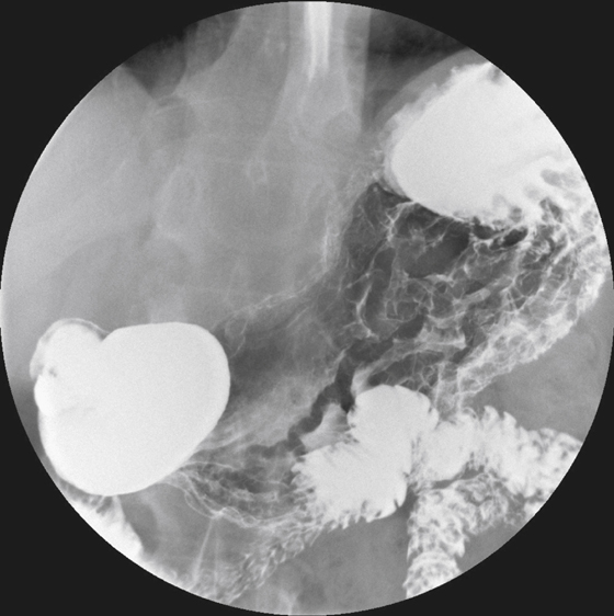
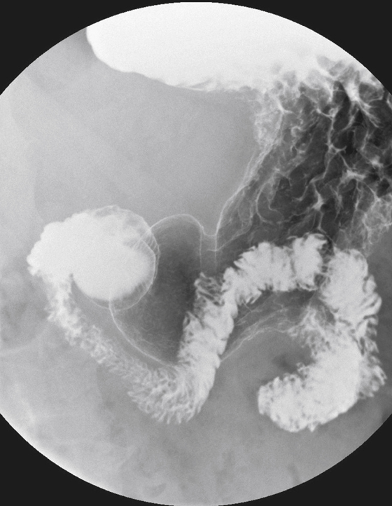
History: A 56-year-old man presents with a long history of heartburn and dyspepsia.
1. Which of the following should be included in the differential diagnosis of the imaging finding shown in the figures? (Choose all that apply.)
2. What neoplasm most commonly produces thickened gastric folds?
3. What causes isolated fundal gastric varices?
4. What is the most common infectious cause of thickened folds?
A. Mycobacterium avium-intracellulare
ANSWERS
CASE 54
Gastric Fold Thickening
1. A, B, C, and E
2. A
3. D
4. D
Reference
Levine MS. Stomach and duodenum: differential diagnosis. In: Gore RM, Levine MS, eds. Textbook of Gastrointestinal Radiology. 2nd ed Philadelphia: WB Saunders; 2000:698–702.
Cross-Reference
Gastrointestinal Imaging: THE REQUISITES, 3rd ed, p 64.
Comment
It is extremely common to find thickened gastric folds on upper gastrointestinal examinations. The width of the gastric folds is usually about 3 to 5 mm in the distal prepyloric stomach and 8 to 10 mm proximally in the fundus. When the stomach is fully distended, normal gastric folds tend to run parallel to the lumen, whereas folds that are irregularly thickened, nodular, or serpiginous in appearance are usually considered abnormal.
Associated gastric wall thickening also may be visible on CT examination.
The presence of thickened rugal folds, gastric wall thickening, or both is a nonspecific finding. A variety of diseases can produce this radiologic finding, which can even be seen in healthy patients. The most common cause is some type of gastritis, such as alcoholic gastritis. H. pylori infection of the stomach is now recognized as an extremely common cause of inflammatory disease of the stomach and is by far the most common infectious process of the upper gastrointestinal tract. Zollinger-Ellison syndrome (gastrinoma) should always be considered, although it is rare. Numerous benign infiltrating processes, including eosinophilic gastritis, sarcoidosis, amyloidosis, Crohn’s disease, and Ménétrier’s disease, also can produce fold thickening. Of the neoplastic processes, lymphoma most typically manifests as thickened folds. Adenocarcinoma and even metastases also must be considered but are less common.
Varices of the stomach usually have associated esophageal varices and are related to increased portal venous pressure resulting from a variety of causes. Isolated fundal gastric varices, however, are a specific condition associated with splenic vein occlusion. The spleen normally drains via the splenic vein, but if this vein occludes, there are short gastric veins that act as collateral circulation in the fundal region of the stomach. These veins course over the proximal stomach and connect to the coronary vein, which then flows into the portal circulation. Splenic vein thrombosis is usually the sequela of pancreatitis or pancreatic carcinoma and less commonly the result of retroperitoneal processes, surgery, or hypercoagulability state.
[/level-membership-for-gastroenterology-and-hepatology-category][not-level-membership-for-gastroenterology-and-hepatology-category]CASE 54
History: A 56-year-old man presents with a long history of heartburn and dyspepsia.
1. Which of the following should be included in the differential diagnosis of the imaging finding shown in the figures? (Choose all that apply.)
2. What neoplasm most commonly produces thickened gastric folds?
3. What causes isolated fundal gastric varices?
[/not-level-membership-for-gastroenterology-and-hepatology-category]
