CASE 54
1. Which chambers are most closely associated with the mass? (Choose all that apply.)
A. Left atrium
C. Right atrium
A. Left atrium
B. Right atrium
D. Aortic root
3. What is the most likely diagnosis?
A. Lipoma
D. Metastasis
A. It represents a metastasis.
B. It represents an angiosarcoma.
ANSWERS
Reference
Maurer AH, Burshteyn M, Adler LP, et al. How to differentiate benign versus malignant cardiac and paracardiac 18F FDG uptake at oncologic PET/CT. Radiographics. 2011;31(5):1287–1305.
Cross-Reference
Cardiac Imaging: The REQUISITES, ed 3, pp 149, 282.
Comment
Clinical Features
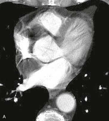
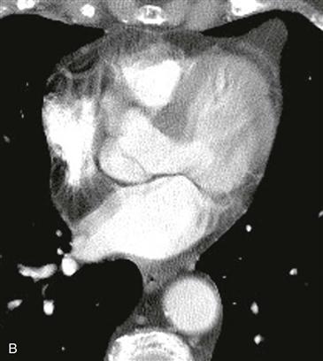
Lipomatous hypertrophy of the interatrial septum is benign and is characterized by fat in the interatrial septum. It occurs more frequently in elderly, obese patients and is associated with an increased amount of epicardial fat. Lipomatous hypertrophy of the interatrial septum is composed of brown fat and can be FDG avid on PET scan. This lesion is within the interatrial septum, not in the atrium, and it does not normally need to be resected.
Imaging
The lesion is sometimes seen on echocardiography or PET scan, and patients are referred to CT or MRI to differentiate intraatrial lipoma or thrombus from lipomatous hypertrophy (Figs. A and B). In other instances, the mass is discovered incidentally on CT or MRI. When MRI is performed, fat saturation images can confirm the composition of the mass. The mass can have a narrow waist in the region of the fossa ovalis.


