CASE 53
History: A patient presents with chest pain.
1. What are potential etiologies of this abnormality? (Choose all that apply.)
C. Infection
D. Trauma
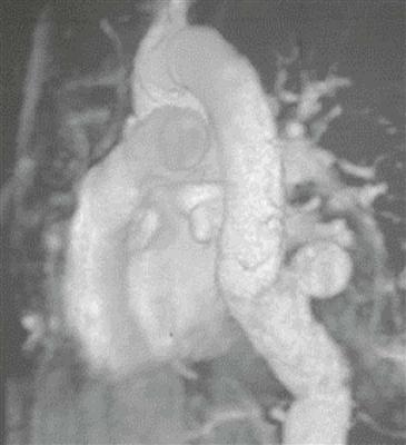
2. Which portion of the aorta is most abnormal?
A. Ascending
B. Arch
C. Descending
D. Abdominal
3. What is the most likely diagnosis?
4. What type of image is shown?
B. Curved multiplanar reformation
ANSWERS
Reference
Reddy GP, Gunn M, Mitsumori LM, et al. Multislice CT and MRI of the thoracic aorta. In: Webb WR, Higgins CB, eds. Thoracic imaging: pulmonary and cardiovascular radiology. ed 2 Philadelphia: Lippincott Williams & Wilkins; 2010.
Cross-Reference
Cardiac Imaging: The REQUISITES, ed 3, pp 377–379.
Comment
Etiology and Pathology
Etiologies of aortic pseudoaneurysm include atherosclerosis (penetrating aortic ulcer), infection, trauma (deceleration injury—although this is an unusual location), and iatrogenic cause. Pseudoaneurysms are characterized by disruption of one or more layers of the vessel wall, whereas true aneurysms have intact walls.
True and False Aneurysm
In a true aneurysm of the aorta, all three layers of the aortic wall are intact. In contrast, a false aneurysm results from a focal disruption of one or more layers of the aortic wall and may be contained by the adventitia and surrounding fibrous tissue.
Imaging
CT and MRI are the best imaging methods for evaluation of a thoracic aortic aneurysm. Disruption of the wall is difficult to identify on imaging. However, many observers use the following rule of thumb: a relatively narrow ostium (<50% of the aneurysm diameter) suggests that the outpouching is a pseudoaneurysm (Figure), and a wide ostium suggests a true aneurysm.


