CASE 52
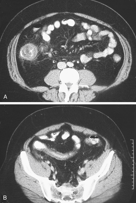
History: A 40-year-old man presents with right lower quadrant abdominal pain and fever.
1. Which of the following should be included in the differential diagnosis of the dominant imaging finding on figure A? (Choose all that apply.)
2. What is the most common clinical manifestation of Crohn’s disease?
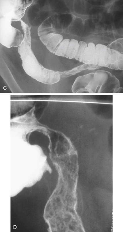
3. Which of the following correctly describes the string sign?
A. Sign on barium small bowel study where the small bowel lumen resembles a string
4. What parts of the gut are most commonly involved with Crohn’s disease?
ANSWERS
CASE 52
Crohn’s Disease of the Terminal Ileum
1. B, C, D, and E
2. D
3. A
4. B
References
Horsthuis K, Bipat S, Bennink RJ, Stoker J. Inflammatory bowel disease diagnosed with US, MR, scintigraphy, and CT: meta-analysis of prospective studies. Radiology. 2008;247:64–79.
Wills JS, Lobis IF, Denstman FJ. Crohn disease: state of the art. Radiology. 1997;202:597–610.
Cross-Reference
Gastrointestinal Imaging: THE REQUISITES, 3rd ed, p 126.
Comment
Crohn’s disease (or regional enteritis), as seen in the distal ileum of this patient (see figures), is one of the major idiopathic inflammatory bowel diseases (ulcerative colitis being the other). It is a transmural granulomatous inflammatory process and has been described in medical literature with various attached names for more than 200 years.
The cardinal clinical presentation includes diarrhea, abdominal pain, and weight loss. The incidence is higher in Europe and North America than in Asia. The incidence has increased steadily in the West until about the mid-1980s, when it began to stabilize. It tends to be a disease of young people (15 to 25 years old), although there is a second much smaller peak in the seventh decade. It has long been thought to be an immunologic response to some stimulating agent, although such an agent has never been identified.
Barium studies can be helpful with colonic involvement by identifying the specific pattern, which is usually quite different from that of ulcerative colitis. However, when only the terminal ileum alone is involved, all imaging studies are much less specific. The presence of fistulous communication, perianal fissuring, and extragastrointestinal manifestation may be helpful.


