CASE 51
1. What shunt lesions can cause pulmonary hypertension?
B. Ventricular septal defect (VSD)
C. Patent ductus arteriosus (PDA)
D. Partial anomalous pulmonary venous connection (PAPVC)
2. What is the most likely diagnosis?
A. ASD
B. VSD
C. PDA
D. PAPVC
3. Which lesion is commonly associated with this anomaly?
A. VSD
B. ASD
C. PDA
4. Which other lesion is most physiologically similar to PAPVC?
A. VSD
B. ASD
C. PDA
ANSWERS
Reference
Reddy GP, Higgins CB. Magnetic resonance imaging of congenital heart disease: evaluation of morphology and function. Semin Roentgenol. 2003;38(4):342–351.
Cross-Reference
Cardiac Imaging: The REQUISITES, ed 3, pp 330–335.
Comment
Physiology and Associated Anomalies
Depending on the severity of the shunt, patients with PAPVC may be asymptomatic; may have dyspnea, cardiac murmur, and decreased exercise tolerance; or may have pulmonary hypertension. Because it is an atrial-level shunt, PAPVC is physiologically similar to ASD. PAPVC is associated with other congenital abnormalities, most commonly a sinus venosus ASD, in which a connection exists between the left atrium and the superior vena cava as the cava enters the right atrium.
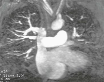
Imaging
Medical and surgical management depends on accurate evaluation of the number and site of anomalous pulmonary veins and the presence of an ASD or other congenital anomaly. Gadolinium-enhanced magnetic resonance angiography (MRA) can accurately depict the presence, location, and size of anomalous veins in patients with PAPVC (Figure). MRI has the advantage over CT and angiography of no ionizing radiation or iodinated contrast agent.


