CASE 50
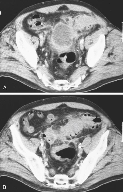
History: A 65-year-old woman presents with 4 weeks of lower abdominal pain, diarrhea, and fever; urine culture growing Proteus mirabilis.
1. Which of the following should be included in the differential diagnosis of the imaging finding shown in figure A? (Choose all that apply.)
2. Regarding colonic diverticula, which of the following statements is false?
A. Colonic diverticula are true diverticula.
B. Colonic diverticula align along the taenia coli.
C. Colonic diverticula are pulsion lesions.
D. Diverticula are most common in the sigmoid segment.
E. The etiology of diverticulitis is similar to that of appendicitis.
3. Which of the following statements regarding colonic diverticular disease is true?
A. Diverticular disease is a common worldwide condition.
B. Most patients present with hemorrhage or diverticulitis.
C. Diverticular disease is related to the modern Western diet high in animal fats.
D. More than 60% of people in the United States older than 60 years have colonic diverticula.
4. Which of the following statements regarding imaging of colonic diverticulitis is false?
A. The observation of narrowing means that there is or has been an episode of diverticulitis.
B. A component of the narrowing is reversible.
C. CT is a useful modality in diagnosing acute diverticulitis.
D. Barium enema can be diagnostic.
ANSWERS
CASE 50
Sigmoid Diverticulitis
1. B, C, D, and E
2. A
3. D
4. A
References
Birnbaum BA, Balthazar EJ. CT of appendicitis and diverticulitis. Radiol Clin North Am. 1994;32:885–898.
Cross-Reference
Gastrointestinal Imaging: THE REQUISITES, 3rd ed, p 302.
Comment
Diverticular disease of the colon (diverticulosis coli) is a common disease of the Western world. The older the patient, the greater the likelihood of having the disease. However, in the last 3 or 4 decades there appears to be an increased incidence in younger patients. Diverticula formation is probably related to increased intraluminal pressures, decreased bowel transit times, and diminishing quantities of fiber in the diet in Western industrialized countries. Most of these patients are asymptomatic or manifest vague abdominal discomfort. A small group experience rectal bleeding, although a massive rectal bleed secondary to diverticulosis is uncommon (less than 5%). Very often the offending diverticulum is in the right colon.
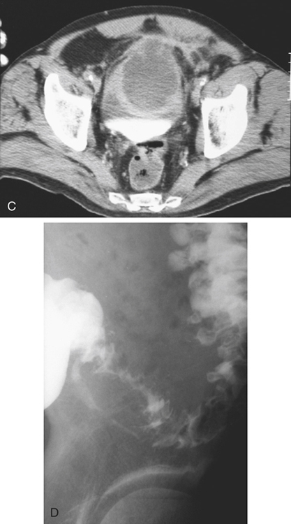
About 15% of this population develops frank diverticulitis, such as shown in the images accompanying this case (see figures). The pathogenesis is thought to be infection secondary to impacted fecal matter within the diverticula, local peridiverticular inflammation, and finally, if untreated, frank diverticulitis with pericolonic abscess formation. Some of the abscesses spontaneously drain via communication with the colonic lumen, such as shown in the barium enema image (see figures). Most do not and will require surgical intervention or radiologic placement of an abscess drainage catheter. It is believed by some that the origin of the so-called giant sigmoid diverticulum is, in fact, the sequela of spontaneous drainage of a large diverticular abscess with the remaining cavity permanent, communicating with the sigmoid colon and epithelialized over time.
There is little doubt that CT of the abdomen is the best imaging method for diverticulitis. Not only can mild pericolic inflammatory changes be detected but also pericolic abscesses.


