CASE 50
1. What should be included in the differential diagnosis for pulmonary edema in an infant? (Choose all that apply.)
D. Total anomalous pulmonary venous connection
2. What is the finding seen on the chest radiograph?
A. Cardiomegaly
3. What is the most likely diagnosis in this patient?
D. Total anomalous pulmonary venous connection
4. Which type of total anomalous pulmonary venous connection is most likely to manifest with a “snowman heart” configuration on chest radiography?
A. Supracardiac
B. Cardiac
C. Infracardiac
D. Mixed
ANSWERS
CASE 50
References
Dillman JR, Yarram SG, Hernandez RJ. Imaging of pulmonary venous developmental anomalies. AJR Am J Roentgenol. 2009;192(5):1272–1285.
Higgins CB. Radiography of congenital heart disease. In: Webb WR, Higgins CB, eds. Thoracic imaging: Pulmonary and cardiovascular radiology. ed 2 Philadelphia: Lippincott Williams & Wilkins; 2010.
Cross-Reference
Cardiac Imaging: The REQUISITES, ed 3, pp 328–330.
Comment
Types of Total Anomalous Pulmonary Venous Connection
In this anomaly, all the pulmonary veins drain into the systemic veins or directly into the right atrium. In the supracardiac type (type I), the enlarged mediastinal veins cause the “snowman heart” appearance. In the cardiac type (type II), the pulmonary veins drain into the coronary sinus or right atrium. In the infracardiac type (type III), the veins connect to the portal vein, hepatic vein, or ductus venosus. Venous flow is obstructed by the passage of the pulmonary veins across the esophageal hiatus, leading to pulmonary congestion and edema without cardiac enlargement.
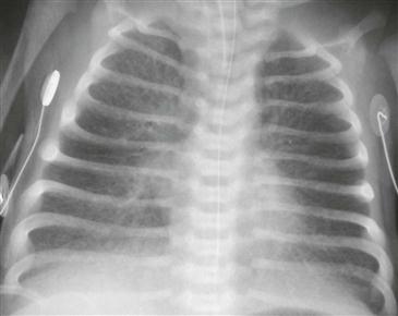
Radiography
The chest radiograph typically shows shunt vascularity. In total anomalous pulmonary venous connection type III (connection below the diaphragm), the radiograph shows pulmonary edema with a normal-sized heart (Figure).


