CASE 5
1. What should be included in the differential diagnosis for a left-to-right shunt? (Choose all that apply.)
A. Partial anomalous pulmonary venous return (PAPVR)
2. What is the cardiac abnormality most commonly associated with this condition?
3. Which syndrome or complex is always associated with this entity?
B. Holt-Oram
C. Scimitar
D. Carney
4. At which pulmonic-to-systemic flow (Qp/Qs) ratio is repair generally recommended?
A. 1
B. 0.3
C. 1.7
D. The Qp/Qs ratio is not used to decide when to repair this lesion
ANSWERS
Reference
Ho ML, Bhalla S, Bierhals A, et al. MDCT of partial anomalous pulmonary venous return (PAPVR) in adults. J Thorac Imaging. 2009;24(2):89–95.
Cross-Reference
Cardiac Imaging: The REQUISITES, ed 3, pp 330–335.
Comment
Epidemiology and Treatment
PAPVR is an uncommon congenital venous anomaly, which occurs in less than 1% of the population. It is a left-to-right shunt with a pulmonary vein or veins bypassing the systemic circulation. When discovered in children, it overwhelmingly affects the right upper lobe (90% of cases) and is highly associated with other cardiac malformations including sinus venosus ASD. Repair of PAPVR is recommended when the Qp/Qs ratio exceeds 1.5 to 2.0. Most children meet these criteria, and usually both ASD and PAPVR are surgically corrected.
Adult Presentation and Management
PAPVR is discovered in adults incidentally on imaging performed for other reasons. It is frequently asymptomatic and most commonly affects the left upper lobe (47%) followed by the right upper lobe (38%). Sinus venosus ASD occurs in 42% of cases of adult right upper lobe PAPVR. Surgery is often not needed in adults because they are asymptomatic.
Imaging
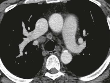
Axial CT shows a right superior pulmonary vein draining directly into the superior vena cava (Figure), consistent with PAPVR. Images more inferior at the level of the upper left atrium (not shown) did not reveal a sinus venosus ASD in this case.