CASE 49
1. What are causes of pacemaker malfunction? (Choose all that apply.)
B. Disconnection of lead from generator
2. What is the most likely diagnosis?
B. Disconnection of lead from generator
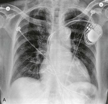
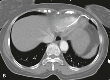
3. Where is the lead that is to the left of midline?
4. Where is the lead that is to the right of midline?
A. Right atrium
C. Left atrium
ANSWERS
Reference
Aguilera AL, Volokhina YV, Fisher KL. Radiography of cardiac conduction devices: a comprehensive review. Radiographics. 2011;31(6):1669–1682.
Comment
Normal Appearance of Pacemakers
Cardiac pacemakers are used in patients with conduction disturbances. Types of pacemakers include single chamber (pacing the right ventricle or right atrium), dual chamber (pacing both the right atrium and right ventricle), and biventricular (pacing the right atrium, right ventricle, and left ventricle). Biventricular pacing is also called cardiac resynchronization therapy. Patients often have combined pacemaker and implantable cardioverter-defibrillators (ICDs). An ICD is used to defibrillate the heart during a potentially fatal arrhythmia and is distinguished from a pacemaker lead by its thick radiopaque shock coil.
Complications of Pacemakers
Acute complications of pacemaker placement include pneumothorax, hemothorax, arrhythmia secondary to inappropriate lead placement, and myocardial perforation. In the setting of long-term pacemaker placement, complications include lead disconnection from the generator, lead fracture, lead displacement, and perforation. Twiddler syndrome is a unique form of lead displacement caused by the conscious or subconscious rotating of the generator in the subcutaneous tissues by the patient. It may be recognized on radiography by noting lead displacement with leads curled around the generator.


