CASE 48
1. What should be included in the differential diagnosis for this patient history? (Choose all that apply.)
A. Arrhythmogenic right ventricular dysplasia
C. Hypertrophic cardiomyopathy (HCM)
2. Based on the images, what is the most likely diagnosis?
A. Arrhythmogenic right ventricular dysplasia
C. HCM
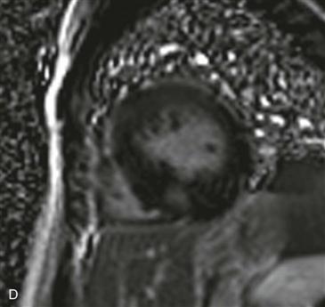
3. What is the distribution of hypertrophy in this patient?
A. Septal
B. Concentric
D. Apical
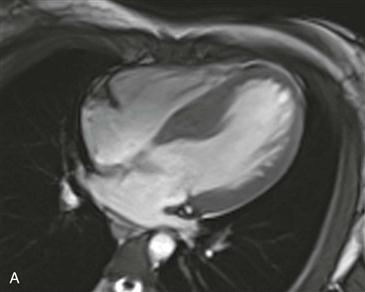
4. Based on Fig. A, which of the following is in the differential diagnosis?
A. Sarcoidosis
B. Angiosarcoma
C. Amyloidosis
ANSWERS
References
Harris SR, Glockner J, Misselt AJ, et al. Cardiac MR imaging of nonischemic cardiomyopathies. Magn Reson Imaging Clin N Am. 2008;16(2):165–183.
Soler R, Rodriguez E, Remuinan C, et al. Magnetic resonance imaging of primary cardiomyopathies. J Comput Assist Tomogr. 2003;27(5):724–734.
Cross-Reference
Cardiac Imaging: The REQUISITES, ed 3, pp 53, 284–288.
Comment
Etiology and Clinical Features
HCM is inherited as an autosomal dominant trait with variable penetrance. Patients have a variable clinical presentation. They may be asymptomatic, or they may have atrial fibrillation, heart failure, syncope, or sudden cardiac death, which is the leading cause of mortality in these patients. Asymmetric hypertrophy of the ventricular septum accounts for 90% of cases of HCM. Other patterns exhibit right ventricular, left ventricular, septal, apical, midventricular, or concentric distribution. Patients with heart failure secondary to significant septal hypertrophy and left ventricular outflow obstruction can be treated with septal myectomy or percutaneous transluminal septal myocardial ablation with ethanol.
Imaging
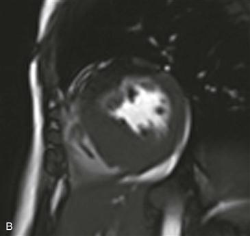
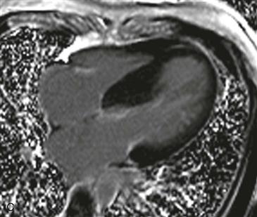
MRI can provide anatomic and functional information in HCM and can be most useful when the diagnosis is in question, when invasive therapy is being considered, or when clinical concern requires more thorough assessment than that provided by echocardiography. MRI can be used to identify the distribution of thickened myocardium, to evaluate for systolic anterior motion of the mitral valve, and to calculate left ventricular mass (Figs. A–D). Late gadolinium enhancement MRI characteristically shows patchy midmyocardial enhancement. To differentiate HCM from a septal neoplasm, gadolinium-chelate contrast agent is administered intravenously. A neoplasm enhances markedly, whereas septal hypertrophic myocardium enhances only slightly. MRI also can be used for functional evaluation of left ventricular outflow tract obstruction and myocardial perfusion and viability.


