CASE 47
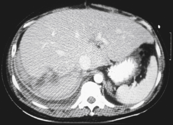
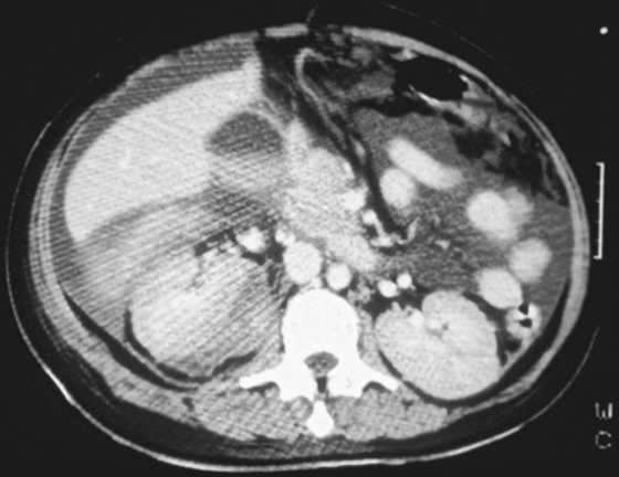
History: A 48-year-old man was involved in a motorcycle accident.
1. Which of the following show signs of a significant injury in the imaging presented? (Choose all that apply.)
2. What injury has the liver suffered?
3. What is the significance of the high density in the fluid adjacent to the liver?
D. Active IV contrast extravasation
4. What is the artifact resulting in the alternating light and dark lines at the left base of the CT images?
D. Scanning beyond the scanning circle
ANSWERS
CASE 47
Liver Laceration
1. B, C, and D
2. A
3. C
4. B
References
Richards JR, Derlet RW. Computed tomography for blunt abdominal trauma in the ED: a prospective study. Am J Emerg Med. 1998;16:338–342.
Cross-Reference
Gastrointestinal Imaging: THE REQUISITES, 3rd ed, p 207.
Comment
After the spleen, the liver is the next most common solid organ injured in blunt trauma to the abdomen. Liver traumas can be solitary, but most often they are seen with multiple injuries of the abdomen, abdominal wall, and thoracic wall as well as musculoskeletal and neurologic injuries elsewhere. By far the most common cause is trauma due to motor vehicle accidents. The injury to the liver is most serious when large blood vessels have been torn, such as the hepatic veins, the portal vessels, or the hepatic artery. Those with hepatic artery tears often do not make it to the emergency department.
Commonly what is seen in CT is a laceration with accompanying intrahepatic or subcapsular hematoma (see figures). Some degree of extrahepatic hematoma is seen in about 80% of cases. Hepatic contusion can manifest as a focal area of diminished density. However, these injuries are almost always self-limiting conditions for which no treatment is required. Survival of patients with intra-abdominal solid organ trauma has increased over the last few decades with improved on-site patient care and rapid transport to a trauma center. Without doubt, a major contributing factor has been the availability of CT imaging in or adjacent to the trauma center.
Blunt liver trauma has been categorized by organ injury scale, including contusion, hematoma, and laceration at one end of the scale, and major vascular injuries and hepatic avulsion at the other.


