CASE 46

History: An 81-year-old man presents with confusion. His family reports also a history of 10 kg weight loss over 6 weeks. Initial investigations identified iron deficiency anemia.
1. Which of the following should be included in the differential diagnosis of the imaging finding shown? (Choose all that apply.)
2. What is the name of the association of gastric cancer metastatic to the left axilla?
3. Which is the most appropriate imaging modality to assess local spread of gastric cancer?
4. Which of the following statements about gastric cancer is true?
A. Gastric cancer is the most common GI malignancy.
B. About 10% of gastric cancers are of the linitis plastic type.
D. Patients with the linitis plastica type of gastric cancer usually present with dysphagia.
ANSWERS
CASE 46
Linitis Plastica of the Stomach
1. A, C, D, and E
2. A
3. A
4. B
References
Iyer R, Dubrow R. Imaging upper gastrointestinal malignancy. Semin Roentgenol. 2006;41(2):105–112.
Cross-Reference
Gastrointestinal Imaging: THE REQUISITES, 3rd ed, pp 57-59.
Comment
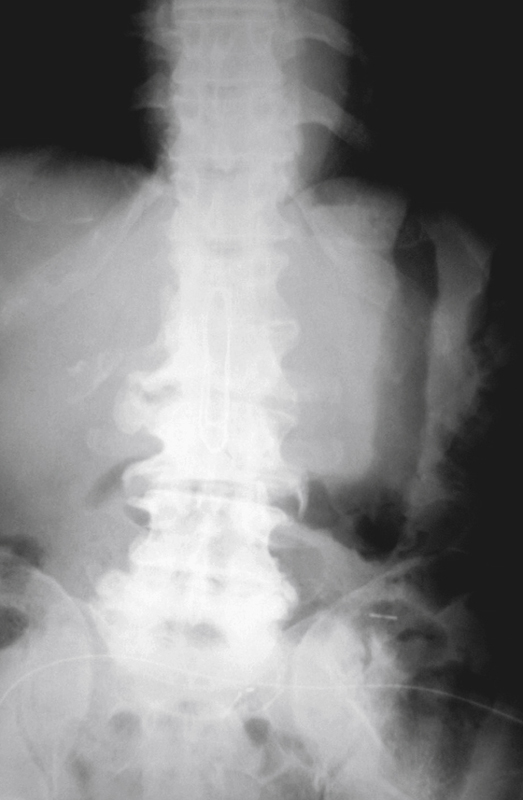
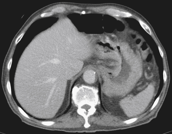
Close attention to the gas pattern of the stomach on plain film images can sometimes reveal diffuse abnormalities of the stomach, such as in this case (see figures). The gas pattern of the stomach in this patient suggests a narrow rigid stomach with loss of pliability, which is confirmed by barium upper GI study (see figures). A CT image shows diffuse thickening of the gastric wall (see figures). In this case, the findings (linitis plastica) are a result of diffuse scirrhous adenocarcinoma of stomach. However, other conditions can also give a similar appearance. Metastatic disease involving the stomach (especially from breast and lung) can result in an identical appearance, as can Hodgkin’s type desmoplastic lymphoma of the stomach and inflammatory diseases such as severe diffuse peptic gastritis, corrosive gastritis, radiation gastritis, sarcoidosis, and Crohn’s disease (ram’s horn stomach), as well as some reported cases of syphilis.


