[level-membership-for-gastroenterology-and-hepatology-category]CASE 45
History: A 67-year-old man with melena. Upper GI endoscopy found an ulcerated extrinsic mass effect on the third part of the duodenum.
1. Which of the following should be included in the differential diagnosis of the case and imaging finding shown? (Choose all that apply.)
2. What is the most common histologic type of pancreatic malignancy?
A. Acinar epithelial adenocarcinoma
B. Ductal epithelial adenocarcinoma
3. What is the most common clinical presentation of a patient with pancreatic cancer?
4. Where are most pancreatic cancers located?
ANSWERS
CASE 45
Pancreatic Carcinoma
1. A, B, C, and D
2. B
3. A
4. A
References
Chezmar JL. Pancreatic neoplasms. In: Gore RM, Levine MS, eds. Textbook of Gastrointestinal Radiology. 2nd ed Philadelphia: WB Saunders; 2000:1796–1811.
Cross-Reference
Gastrointestinal Imaging: THE REQUISITES, 3rd ed, pp 159-165.
Comment
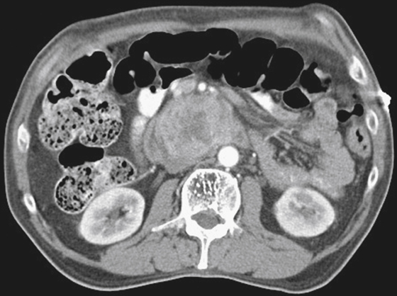
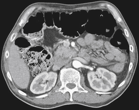
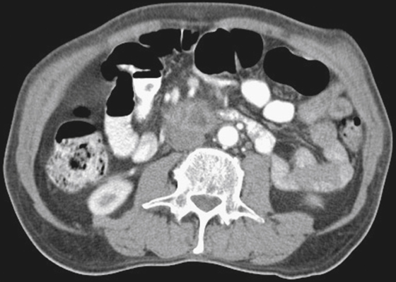
The CT images show a mixed-density pancreatic mass within the head and uncinate process. The adjacent superior mesenteric artery, superior mesenteric vein, and aorta remain separated by a fat plane, suggesting the tumor may be an operable lesion.
The clinical findings of jaundice and painless gallbladder enlargement (Courvoisier’s gallbladder) are associated with this disease, as is Trousseau’s syndrome (venous thrombotic disease) and occasionally new onset of diabetes mellitus. At the time of diagnosis, patients with symptoms have a median survival rate of about 20 months.
On occasion, and usually as an unexpected incidental finding, one can come across a small pancreatic head lesion, often in the uncinate process, that is asymptomatic. These fortunate patients may be candidates for resections and a Whipple procedure with an increased life expectancy.
[/level-membership-for-gastroenterology-and-hepatology-category][not-level-membership-for-gastroenterology-and-hepatology-category]CASE 45
History: A 67-year-old man with melena. Upper GI endoscopy found an ulcerated extrinsic mass effect on the third part of the duodenum.
1. Which of the following should be included in the differential diagnosis of the case and imaging finding shown? (Choose all that apply.)
2. What is the most common histologic type of pancreatic malignancy?
A. Acinar epithelial adenocarcinoma
B. Ductal epithelial adenocarcinoma
[/not-level-membership-for-gastroenterology-and-hepatology-category]
