CASE 45
History: A patient presents with fever.
1. What should be included in the differential diagnosis based on the radiograph? (Choose all that apply.)
A. Lymphoma
2. If the mass has a density of 8 HU, what is the most likely diagnosis?
A. Thymoma
C. Thymic cyst
D. Lymphoma
3. What is the most common location of a pericardial cyst?
C. Superior pericardial recess
D. Subcarinal
4. Which of the following is least specific for a pericardial cyst?
A. Hounsfield number of 10 on a non–contrast-enhanced CT image
B. Hounsfield number of 30 on both a non–contrast-enhanced CT image and a contrast-enhanced CT image
ANSWERS
References
Kim JS, Kim HH, Yoon Y. Imaging of pericardial diseases. Clin Radiol. 2007;62(7):626–631.
Wang ZJ, Reddy GP, Gotway MB, et al. CT and MR imaging of pericardial disease. Radiographics. 2003;23(Spec No):S167–S180.
Cross-Reference
Cardiac Imaging: The REQUISITES, ed 3, pp 78–79.
Comment
Etiology and Pathology
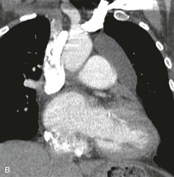
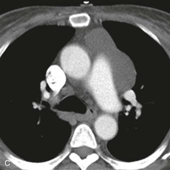
A pericardial cyst is a benign developmental lesion that is formed when a portion of the embryonic pericardium is pinched off and isolated. It has a thin wall, contains clear fluid, and is well circumscribed. The two most common locations are the right and left cardiophrenic angles. When a pericardial cyst is located in another area of the mediastinum, it can be difficult to differentiate from a bronchogenic, esophageal duplication, neuroenteric, or thymic cyst.
Imaging Features and Diagnosis
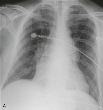
Radiographs typically show a nonspecific mediastinal contour abnormality (Fig. A). On CT and MRI, pericardial cysts are round or ovoid and are contiguous with the normal pericardium (Fig. B). CT shows a well-circumscribed mass (Fig. C). The density may be in the range of simple fluid, in which case the diagnosis is straightforward. If the density is higher, non–contrast-enhanced and contrast-enhanced CT can be performed to assess for enhancement. Pericardial cysts do not enhance, in contrast to neoplasms. Alternatively, MRI can be performed. On MRI, these lesions typically exhibit signal characteristics consistent with simple cysts found elsewhere in the body.


