Procedure 43 Carpal Wedge Osteotomy for Congenital Wrist Flexion Contracture (Arthrogryposis)
 See Video 35: Triceps Lengthening and Elbow Release in Arthrogryposis

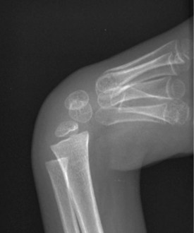
Examination/Imaging


Positioning


Exposures
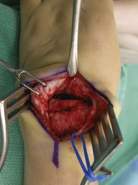
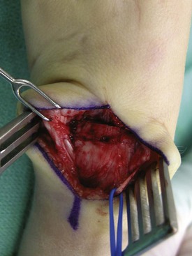
 A single 4-cm longitudinal incision centered over the dorsum of the wrist is made. The extensor retinaculum is identified, and skin flaps are raised on both sides superficial to the retinaculum.


Procedure
Step 1
 A wedge of bone is removed from the midcarpal joint using an osteotomy. The amount of bone removed depends on the preoperative flexion and ulnar deviation deformity. The proximal cut is made perpendicular to the long axis of the radius, and the distal cut is made perpendicular to the metacarpals. This results in a wedge because of the flexion deformity. The wedge should be made wider on the radial side if correction of an ulnar deviation deformity is also required (Fig. 43-4).
Step 1 Pearls
Intraoperative fluoroscopy can be used to confirm the position of the osteotomy in the older child. In the younger child whose carpus has not yet ossified, the position should be confirmed visually.
A scalpel blade can be used in the younger child because the carpus is relatively soft. An osteotome will be required in the older child. Using a saw is not necessary because there is less control of the cut and the bone is soft.
Step 2
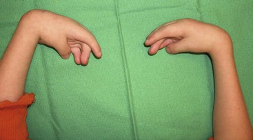
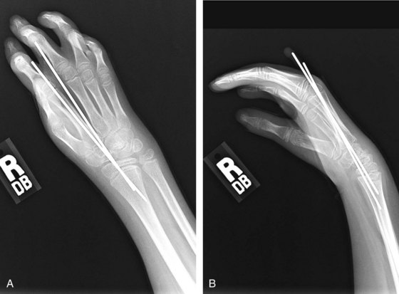
 The aim of the wedge osteotomy is to get the wrist into a posture of slight extension (Fig. 43-5). If the wrist cannot be brought into this position, tendons on the volar side need to be released. A 4-cm volar longitudinal incision is made proximal to the wrist crease and ulnar to the palmaris longus.


Postoperative Care and Expected Outcomes
 The limb is immobilized in an above-elbow fiberglass cast. The cast will be maintained for 6 weeks to allow the osteotomy site to heal. The K-wires are removed at 6 weeks, and the osteotomy site is protected with use of an intermittent splint for 3 months. Finger motion is permitted within the cast.
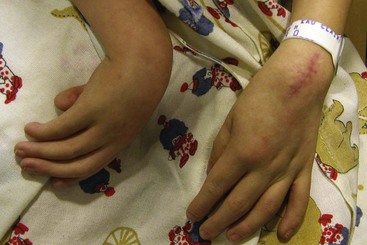
 Formal therapy is usually not required in younger children who adjust to the repositioned wrist. Surgery reliably repositions the wrist, which improves grasp (Fig. 43-7). Improvements in activities of daily living are dependent on degree of limb involvement and overall status of the child.