CASE 43
1. Which cardiac chambers are abnormal? (Choose all that apply.)
A. Left atrium
C. Right atrium
2. What is the most likely diagnosis?
A. Thrombus
B. Myxoma
C. Metastasis
D. Angiosarcoma
3. What is the most likely etiology of the abnormality?
A. Arrhythmia
C. Aneurysm
4. What is the most appropriate way to establish the diagnosis?
B. MRI
C. Angiography
D. Biopsy
ANSWERS
References
Chung JH, Mitsumori LM, Ordovas KG, et al. Heart as a source of stroke: imaging evaluation with computed tomography. J Thorac Imaging. 2012;27(3):W52–W60.
Tatli S, Lipton MJ. CT for intracardiac thrombi and tumors. Int J Cardiovasc Imaging. 2005;21(1):115–131.
Cross-Reference
Cardiac Imaging: The REQUISITES, ed 3, p 274.
Comment
Cardiac Masses
Thrombus is the most common cardiac or paracardiac mass. Among cardiac and paracardiac neoplasms, secondary tumors occur at 40 times the rate of primary neoplasms. Secondary tumors can involve the heart by direct extension (most commonly lymphoma or lymphadenopathy metastatic from lung or breast carcinoma) or by hematogenous spread (most commonly lung or breast carcinoma or melanoma). Primary benign tumors of the heart include myxoma, lipoma, and rhabdomyoma (associated with tuberous sclerosis). Primary malignant neoplasms include angiosarcoma and rhabdomyosarcoma.
Etiologies
Thrombi in the left ventricle are most commonly associated with an aneurysm. Coagulation disorders and flow abnormalities are less common causes of left ventricular thrombus.
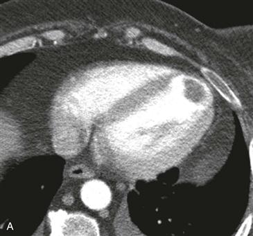
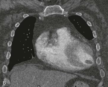
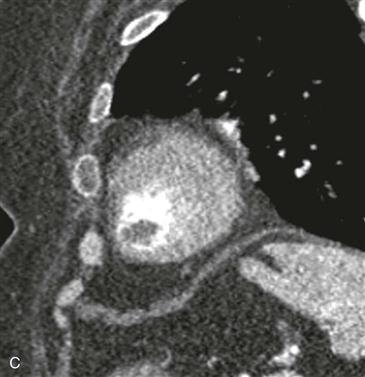
Imaging Findings and Diagnostic Criteria
Echocardiography and CT can identify cardiac masses (Figs. A–C). MRI is the most accurate imaging examination for differentiation of tumor from thrombus. Neoplasms enhance homogeneously or heterogeneously after administration of gadolinium-chelate contrast agent, whereas bland thrombi typically do not enhance except in the periphery. On steady-state free precession (SSFP) MRI, a tumor usually shows intermediate signal intensity, and thrombus tends to demonstrate low signal intensity. However, myxomas can contain calcium and iron and can be dark on SSFP images.


