CASE 42
History: A patient presents with wheezing.
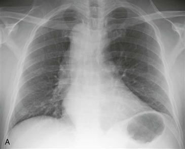
1. What should be included in the differential diagnosis based on the posteroanterior radiograph? (Choose all that apply.)
C. Right aortic arch with aberrant left subclavian artery
D. Mirror-image right aortic arch
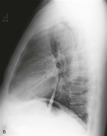
2. Based on the posteroanterior and lateral radiographs, what is the most likely diagnosis?
C. Right aortic arch with aberrant left subclavian artery
D. Mirror-image right aortic arch
3. What is the clinical significance of this anomaly?
A. Most patients have cyanotic congenital heart disease.
B. It is associated with coarctation of the aorta.
C. It causes subclavian steal syndrome.
4. In this anomaly, what completes the ring on the left side?
ANSWERS
References
Reddy GP, Higgins CB. Magnetic resonance imaging of congenital heart disease: evaluation of morphology and function. Semin Roentgenol. 2003;38(4):342–351.
Stojanovska J, Cascade PN, Chong S, et al. Embryology and imaging review of aortic arch anomalies. J Thorac Imaging. 2012;27(2):73–84.
Cross-Reference
Cardiac Imaging: The REQUISITES, ed 3, pp 413–414.
Comment
Clinical Features
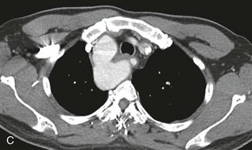
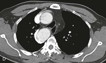
A right aortic arch with an aberrant left subclavian artery is a congenital vascular ring (Figs. A–D). The ring is completed by the left-sided ligamentum arteriosum. Because there is a ring around the trachea and esophagus, these two structures are compressed to a variable extent. The most common symptoms are wheezing, dyspnea, and dysphagia, and patients often exhibit these symptoms during early childhood. The retroesophageal aberrant left subclavian artery may originate from a dilation, known as a diverticulum of Kommerell (Fig. C), which tends to exacerbate the compression on the trachea and esophagus. This type of right aortic arch is weakly associated (5% to 10%) with congenital heart disease, in contrast to the strong association of a mirror-image right arch (>95%).


