CASE 41
History: An 80-year-old woman presents with left-sided abdominal pain and vomiting.
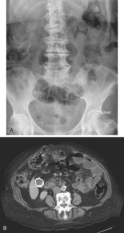
1. What is your differential for the location and diagnosis of the large calcification seen in the right upper quadrant of the abdomen in figure A? (Choose all that apply.)
A. Adrenal gland; e.g., tuberculosis
B. Gallbladder; e.g., porcelain gallbladder
D. Kidney; e.g., calcified cyst
E. Abdominal wall; e.g., old hematoma
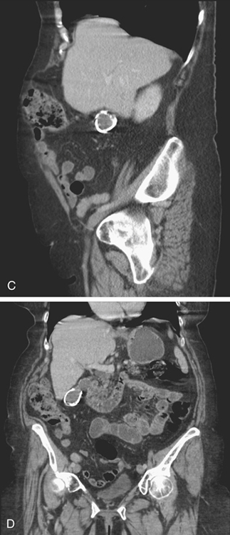
2. Based on the figures, which of the following is the most likely diagnosis?
E. Abdominal wall, old hematoma
3. What is the usual etiologic basis for this condition?
4. Name the major complication associated with this finding.
D. Emphysematous cholecystitis
ANSWERS
CASE 41
Porcelain Gallbladder
1. B, C, D, and E
2. B
3. C
4. C
References
Kane RA, Jacobs R, Katz J, Costello P. Porcelain gallbladder: ultrasound and CT appearance. Radiology. 1984;152:137–141.
Cross-Reference
Gastrointestinal Imaging: THE REQUISITES, 3rd ed, p 254.
Comment
It is thought that chronic cystic duct obstruction and subacute inflammation are the basis of gallbladder wall calcification. Quite often the calcified gallbladder is small and contracted (unlike the example in this case), and it is easy to confuse the finding with calcified gallstones (see figures). However, the porcelain gallbladder carries its own inherent risk, which much outweighs gallstones. The risk of developing carcinoma of the gallbladder in an untreated porcelain gallbladder is high, somewhere between 10% and 30%. Plain film and ultrasound can confuse calcified gallstones and a porcelain gallbladder. However, multidetector CT is much more adept at distinguishing the differences (see figures) and can show the cystic duct obstruction as well.


