Case 4
Baseline 12-Lead ECG
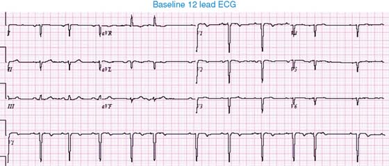
The baseline ECG demonstrates an abnormal P-wave axis, premature supraventricular complexes, and right-axis deviation of the QRS complexes. There is abnormal R wave progression in the precordial leads. The constellation of findings is suggestive of dextrocardia.
Raw Planar Images
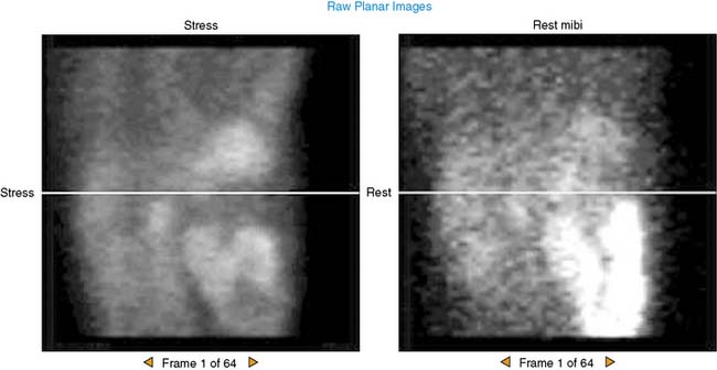
The images were acquired over 180 degrees from the 45-degree right posterior oblique to 45-degree left anterior oblique orientations. Note that the heart is located in the right side of the chest, consistent with dextrocardia. The liver is located on the left, consistent with situs inversus.
SPECT Tomographic Slices
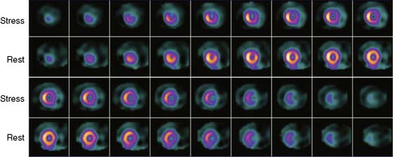
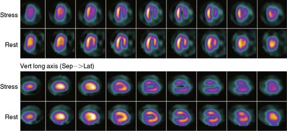
SPECT images are displayed in the standard short-axis, horizontal long-axis, and vertical long-axis orientations. Note that the left and right ventricles are reversed from their usual locations. The appearance is consistent with dextrocardia. Severe inducible ischemia is also present.
Polar Maps
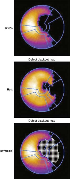
The polar maps suggest that the reversible perfusion defect is located in the anterolateral region, and the perfusion defect corresponds to the territory of the left circumflex coronary artery. Actually, the defect is located in the anteroseptal region and is due to left anterior descending disease. The polar maps are displayed in their normal format and are not corrected for the patient’s dextrocardia.
Poststress Gated SPECT MPI
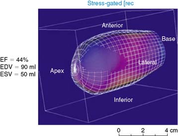
The poststress gated SPECT images demonstrate moderately reduced myocardial systolic thickening in the anteroseptal region, with a computed LVEF of 44%. The regional hypokinesis and reduced LVEF may be due to postischemic myocardial stunning or an imaging artifact created by the severe perfusion defect (leading to an inaccurate tracking of the endocardial border by the automated gated SPECT algorithm).
Cardiac Catheterization
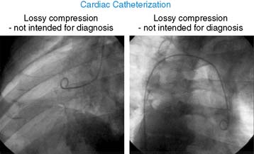
At cardiac catheterization, the heart and aorta demonstrated mirror-image reversal of the normal left-right orientation, consistent with dextrocardia.
Coronary Angiography
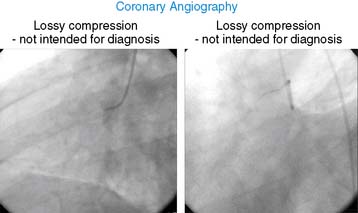
As expected, coronary angiography demonstrated severe LAD stenosis, which was treated with PCI.


