CASE 38
History: A 76-year-old man presents with hemoptysis and hematemesis.
1. Which of the following should be included in the differential diagnosis of the imaging finding shown in figure A? (Choose all that apply.)
2. Which of the following best describes a bull’s-eye lesion?
A. A rounded filling defect with a barium collection at its center owing to ulceration
B. A round metastatic tumor deposit that is black owing to its melanoma origin
C. A region of the stomach that is constricted, inelastic, and rigid
D. A flat lobulated lesion with a reticular mucosal surface pattern
3. What is the most commonly encountered primary tumor resulting in hematogenous metastatic lesions to the stomach?
4. What is the most common site of melanoma metastasis to the gastrointestinal tract?
ANSWERS
CASE 38
Metastatic Lesions to the Stomach
1. B, C, D, and E
2. A
3. D
4. C
References
McDermott VG, Low VHS, Keogan MT, et al: Malignant melanoma metastatic to the gastrointestinal tract. Am J Roentgenol. 1996;166:809–813.
Cross-Reference
Gastrointestinal Imaging: THE REQUISITES, 3rd ed, p 74.
Comment
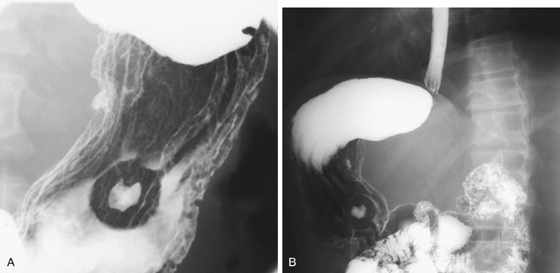
Metastatic lesions to the stomach can take several forms. The form shown in this case is the classic bull’s eye lesion; so named for the appearance of a rounded filling defect with a barium collection at its center. The mound is the metastatic mass arising from the intramural layer of the stomach and the displacement of barium. The bull’s eye is the ulceration and resultant barium collection.
In this case the primary lesion is small cell carcinoma of the lung. However, hematogenous metastatic spread from other lesions can also cause a similar appearance. These lesions include breast cancer and melanoma. In addition, a smaller number of bull’s eye lesions of the stomach can occur as a result of primary gastric tumors. These tumors include some presentations of lymphoma and Kaposi sarcoma. In recent decades the most common cause of bull’s eye lesions in the stomach has been Kaposi’s sarcoma seen in AIDS patients.
Another benign cause of a solitary bull’s eye lesion of the stomach is a gastrointestinal stromal tumor (GIST) in which the mucosa over the center of the intramural lesion ulcerates. Occasionally bull’s eye lesions are seen in the small bowel, but this location is uncommon.


