CASE 38
History: A patient presents with chest pain.
1. Which letters of the alphabet can be used to describe the configuration of the calcification? (Choose all that apply.)
A. C
B. J
C. L
D. S
E. O
2. Which structure is calcified?
A. Aortic valve
C. Mitral valve
3. What is the most common cause of this finding?
B. Endocarditis
C. Degenerative
4. What valvular lesion is most commonly associated with this finding?
ANSWERS
Reference
Gowda RM, Boxt LM. Calcifications of the heart. Radiol Clin North Am. 2004;42(3):603–617.
Cross-Reference
Cardiac Imaging: The REQUISITES, ed 3, pp 8–9.
Comment
Pathology and Etiology
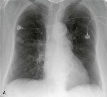
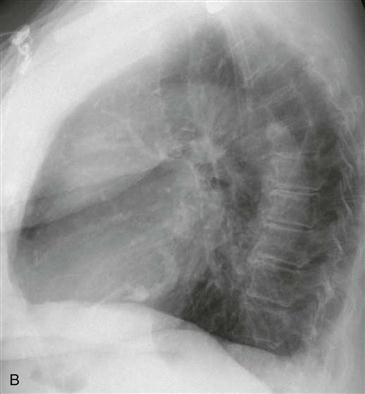
Mitral annular calcification is typically degenerative and related to aging. It occurs more frequently in women and in patients with chronic renal failure. Mitral annular calcification rarely causes symptoms, but when the calcification is extensive, it can lead to mitral regurgitation (or rarely mitral stenosis). Calcification of the mitral valve leaflets themselves is strongly associated with valvular stenosis, most commonly secondary to rheumatic disease.
Imaging Findings and Diagnostic Criteria
Radiographs show calcification of the mitral annulus in a “J,” reverse “C,” or “O” shape (Figs. A and B). It is important to differentiate mitral annular calcification from other calcified cardiac structures on chest radiography. Aortic valve calcification is more anterior than mitral annular calcification and when seen on chest radiography usually indicates at least moderate or severe valvular stenosis. Pericardial calcification is often localized to the atrioventricular groove or can circumferentially surround the heart. It is associated with tuberculosis pericarditis, uremic pericarditis, and radiation therapy and may indicate constrictive pericarditis in the appropriate clinical context. Rarely, intracardiac tumors such as myxoma can calcify diffusely or peripherally.


