CASE 37
History: A child presents with cyanosis.
1. What should be included in the differential diagnosis? (Choose all that apply.)
D. Pulmonary atresia with ventricular septal defect
2. What is the most likely diagnosis?
D. Pulmonary atresia with ventricular septal defect
3. Tetralogy of Fallot comprises all of the following except:
C. Pulmonary infundibular stenosis
E. Right ventricular hypertrophy
4. Which of the following is a severe variant of tetralogy of Fallot?
ANSWERS
Reference
Higgins CB. Radiography of congenital heart disease. In: Webb WR, Higgins CB, eds. Thoracic Imaging: Pulmonary and Cardiovascular Radiology. ed 2 Philadelphia: Lippincott Williams & Wilkins; 2010;742–767.
Cross-Reference
Cardiac Imaging: The REQUISITES, ed 3, pp 359–367.
Comment
Tetralogy of Fallot Overview
Tetralogy of Fallot is the most common cyanotic congenital heart disease in children and adults. The lesions of tetralogy of Fallot are ventricular septal defect, pulmonary infundibular stenosis, right ventricular hypertrophy, and overriding aorta. Most patients with tetralogy of Fallot have decreased pulmonary vascularity, but vascularity can be normal if the infundibular stenosis is mild. Approximately 25% of patients have a right aortic arch. Pulmonary stenosis can occur at multiple levels, including subvalvular or infundibular (most common), valvular, supravalvular, and peripheral.
Pulmonary Atresia with Ventricular Septal Defect
Pulmonary atresia with ventricular septal defect is a severe variant of tetralogy of Fallot in which the pulmonary valve is atretic. Blood reaches the lungs via systemic-to-pulmonary collateral vessels.
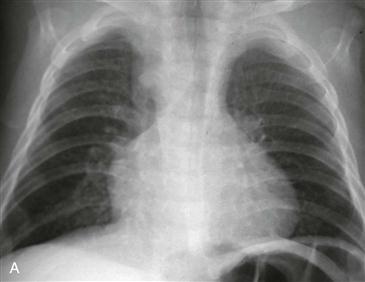
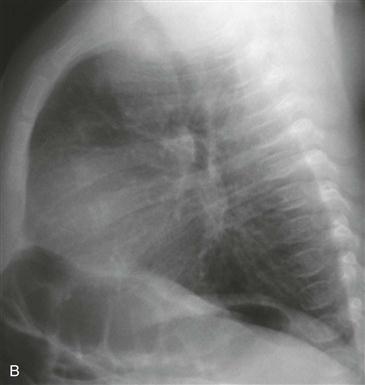
Imaging
In tetralogy of Fallot, radiographs typically demonstrate decreased vascularity, concavity of the pulmonary artery segment, and sometimes an upturned cardiac apex. Chest radiograph in an infant typically demonstrates normal to decreased pulmonary vascularity, normal heart size, and a right aortic arch (Figs. A and B). Echocardiography and angiography are usually performed preoperatively. MRI and CT are occasonally performed preoperatively. MRI is often performed after surgical repair to monitor right ventricular function and to identify and quantify pulmonary regurgitation, which is a complication of surgery.


