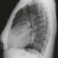
CASE 34
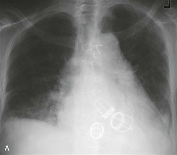
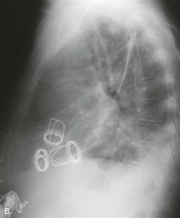
1. Which valves have been replaced? (Choose all that apply.)
A. Aortic
B. Mitral
C. Pulmonary
D. Tricuspid
2. What is the most common cause of multivalve disease?
3. What is the primary advantage of a tissue valve prosthesis over a mechanical valve prosthesis?
A. Safer in the MRI environment
B. No need for anticoagulation
C. Greater durability of valve
D. Less likelihood of calcification
4. Which MRI technique could be used to quantify regurgitation of a prosthetic valve?
ANSWERS
References
Steiner RM, Mintz G, Morse D, et al. The radiology of cardiac valve prostheses. Radiographics. 1988;8(2):277–298.
Walker CM, Reddy GP, Steiner RM. Radiology of the heart. In: Rosendorff C, ed. Essential Cardiology. ed 3 New York: Springer; 2013.
MRIsafety.com. http://www.mrisafety.com/. Accessed February 14, 2013.
Cross-Reference
Cardiac Imaging: The REQUISITES, ed 3, pp 49–50.
Comment
Etiology and Treatment
Rheumatic heart disease is the most common cause of three-valve disease. Patients with three-valve disease may present with heart failure and severe cardiomegaly. Surgical correction is complex, and the mortality rate is approximately 5%.
Types of Prosthetic Valves
Typically, prosthetic mitral and aortic valves are mechanical, and a tricuspid prosthesis may be mechanical or a tissue valve (bioprosthesis). A bioprosthesis can be a heterograft (porcine), a homograft (from a cadaver), or an autograft, in which the patient’s own pulmonary valve and root are used to replace the aortic valve and root. When an autograft is used, a homograft is placed into the pulmonary position.